Zygomatic reduction surgery is intended to reduce the width of the face when the contour of the middle face appears somewhat prominent, while also gently improving the contour of the anterior cheekbone that protrudes below the eyes.
The purpose of this zygomatic reduction surgery can be said to be to achieve a younger-looking, three-dimensional facial shape.
However, after zygomatic reduction surgery, it is sometimes possible to see results in which the face looks flatter and the cheeks appear saggy.
I would like to explain what the cause of such results is.
When consulting with people who have been considering zygomatic reduction surgery,
almost everyone asks the same,
the most "typical" question,
is about "cheek sagging."
What, then, are the causes of cheek sagging associated with zygomatic surgery,
and is cheek sagging inevitable during zygomatic surgery?
I would like to explain these points.
Causes of Cheek Sagging Associated with Zygomatic Surgery
The causes of cheek sagging associated with zygomatic surgery can be broadly considered in three ways.
-
Excessive dissection of the soft tissue from the bone during zygomatic surgery
-
Failure to fix the bone after osteotomy during zygomatic surgery, causing the bone itself to sag downward
-
Loss of the attachment site of the retaining ligament due to the method of cutting the zygomatic body into a trapezoid shape
Among the three causes above,
- Excessive dissection of the soft tissue from the bone during zygomatic surgery is not a major error for surgeons who have accumulated a certain level of experience in zygomatic reduction surgery.
Even with a moderate amount of experience, it is a natural process to perform only the minimum dissection necessary to refine the bone.
Therefore, this cause does not play a major role.
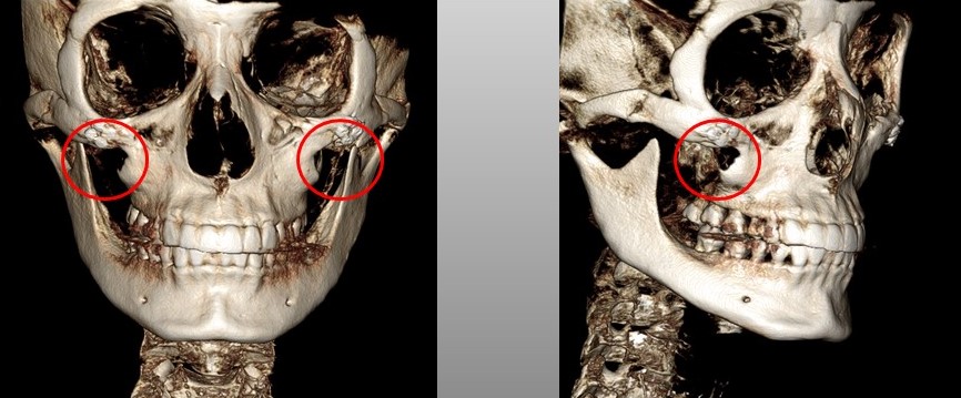
- A case in which the bone itself sags downward because fixation is not performed after osteotomy during zygomatic surgery is as follows.
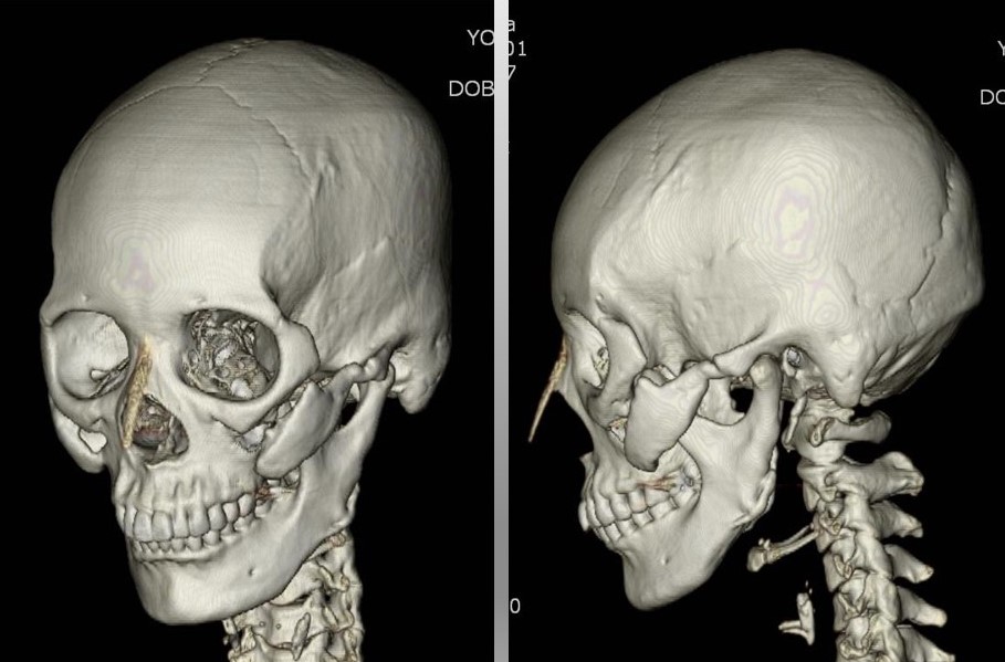
In the 3D CT image above, you can see that a gap has opened at the osteotomy site and nonunion has occurred. You can also see that the bone itself has sagged downward.
Lastly, the third cause,
- Loss of the attachment site of the retaining ligament due to the method of cutting the zygomatic body into a trapezoid shape is the most common cause.
The zygomatic reduction surgery method that is still most commonly performed—cutting the zygomatic body into a trapezoid shape and then completely osteotomizing the zygomatic arch followed by double fixation—is easy to learn and easy to standardize, which is why it can be said to be the most frequently performed method.
Therefore, cheek sagging associated with zygomatic surgery also occurs most frequently.
I think that a somewhat technical and lengthy explanation is needed to understand this.
Because the content may be a little difficult, I will explain it step by step and also attach an explanatory video.
To understand the loss of the retaining ligament attachment site that comes from cutting the zygomatic body into a trapezoid shape, it is first necessary to understand the retaining ligament.
The retaining ligament is a structure in the human body called a Retaining Ligament, and it serves to fix soft tissue to bone or to deeper soft tissue.
Without such retaining ligaments, the bone and the soft tissue (flesh) covering the bone would move independently.
Now, let’s look at where the retaining ligaments are located.
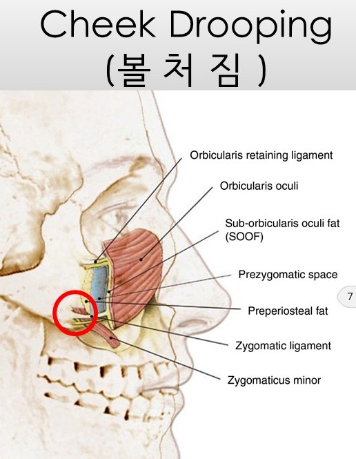
The area marked with a red circle in the image above is the zygomatic retaining ligament among the retaining ligaments.
Zygomatic Ligament
Then what happens when the zygomatic body is cut into a trapezoid shape?
Let’s look at which part of the bone disappears when the zygomatic body is cut into a trapezoid shape, through a schematic diagram.
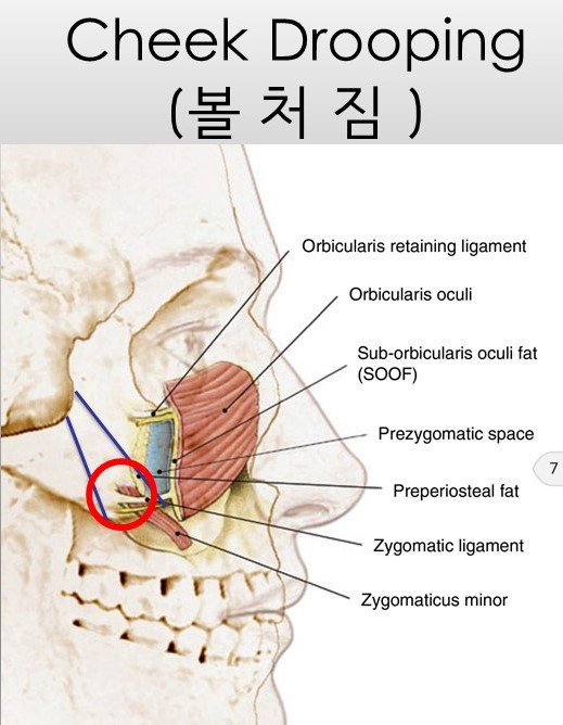
The part marked in blue is the trapezoidal resection area.
In the image above, the blue gradient area is the region that is resected in a trapezoid shape.
As shown in the schematic above, when the zygomatic body is resected in a trapezoid shape, it can be seen that part of the bone to which the retaining ligament attaches is removed.
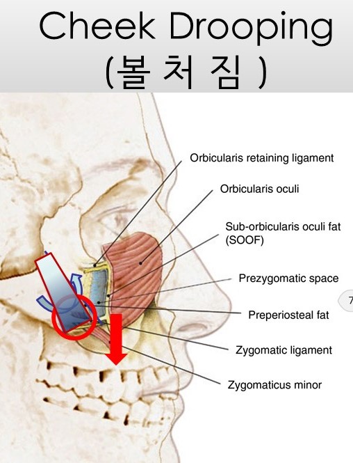
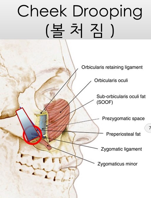
Cheek sagging caused by loss of the retaining ligament attachment site
As shown in the image above, after trapezoidal resection, the loss of the retaining ligament attachment site
causes the soft tissue that had been supported by the retaining ligament to sag.
When the zygomatic body is cut into a trapezoid shape, the lost zygomatic body (red circle) can be observed.
Let’s take a deeper look at what changes may occur when the zygomatic body is cut into a trapezoid shape and the outer zygomatic bone is fixed upward.
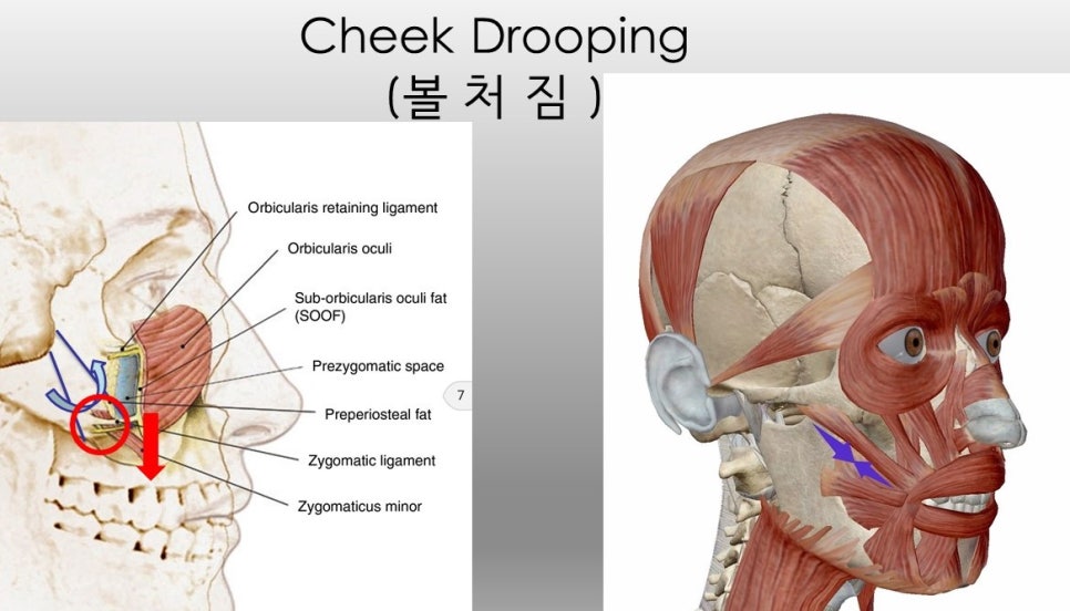
Direction of action of the muscles connected to the zygoma (Zygomaticus major & minor) and the location of the retaining ligament
As shown in the schematic above, when the zygoma is fixed upward, the deep muscles connected to the zygoma pull upward, but the soft tissue covering those muscles sags, and this allows us to understand the mechanism by which the nasolabial fold becomes deeper due to a relative vector.
https://youtu.be/5wmTahdpp5w
Many people ask me.
"They say that cheek sagging inevitably accompanies zygomatic surgery."
Then I answer like this.
"It can vary depending on the surgical method."
They ask me again.
"I heard that zygomatic surgery should be performed through a scalp incision to minimize cheek sagging?"
I answer.
"In fact, among my zygomatic revision surgeries, the satisfaction is said to be highest in patients for whom I performed revision surgery after insufficient results from zygomatic surgery through a scalp incision.