Zygoma reduction surgery can be said to aim to reduce the width of the face when the contour of the middle part of the face appears somewhat prominent, while also gently improving the contour of the front cheekbone that protrudes below the eyes.
When considering this type of zygoma reduction surgery, a precise understanding of the positional movement of the zygomatic arch, which determines the contour of the side cheekbone, is necessary in order to refine the facial outline from the front and make it as narrow as possible.
Recently, interest in posterior zygoma reduction or back cheekbone reduction has gradually increased, but I feel that misunderstandings about back cheekbone reduction are widespread, so I would like to explain them.
The cause of these misunderstandings is that I recently learned that some plastic surgery clinics sometimes disguise the difference in viewing angles between pre- and post-operative CT scans, or use methods that are not understandable from the pre- and post-operative CTs, and I would like to provide a detailed explanation.
In addition, if I were to point out the most important part of posterior zygoma reduction surgery, it would be that although it is important to osteotomize and move the most posterior part of the zygomatic arch, even more important is that the functional structures that form the temporomandibular joint must never be damaged.
To reduce the back cheekbone, if damage is caused to the articular tubercle, which forms the temporomandibular joint, there is also a risk of causing permanent and serious sequelae.
The shape of the zygoma must be approached from a three-dimensional perspective, and the most important thing is to recognize changes in position by setting reference points for each anatomical structure from various angles from which the bone is viewed.
In fact, the zygoma has the following structure.
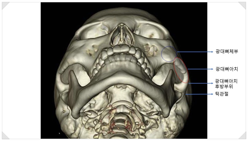
The zygoma, called the zygomatic complex, consists of the zygomatic body and the zygomatic arch.
Zygomatic complex = zygomatic body + zygomatic arch
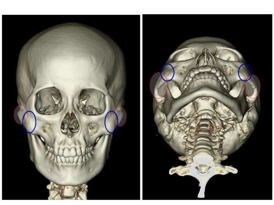
Among these, the area that determines the contour of the side cheekbone and the posterior cheekbone is the zygomatic arch.
The zygomatic arch is structured so that it begins in the front and spreads outward as it goes backward.
In other words, the widest part of the face can be said to be the posterior portion of the zygomatic arch, immediately in front of the temporomandibular joint.
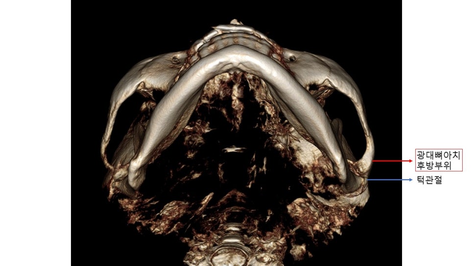
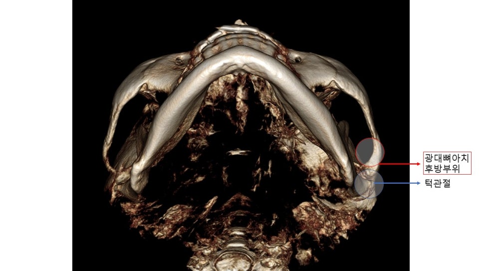
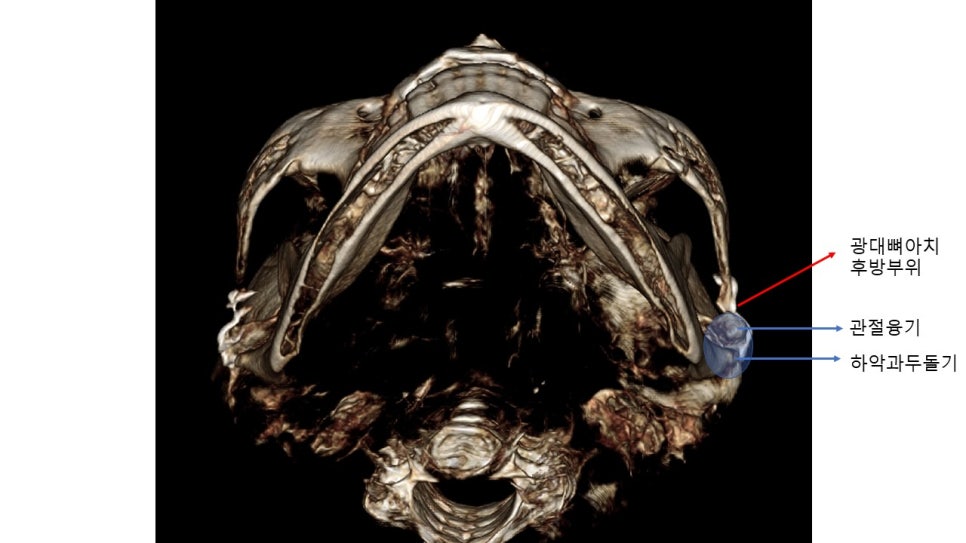
From the angle of looking up at the face, the different shapes of the zygoma appear as follows.
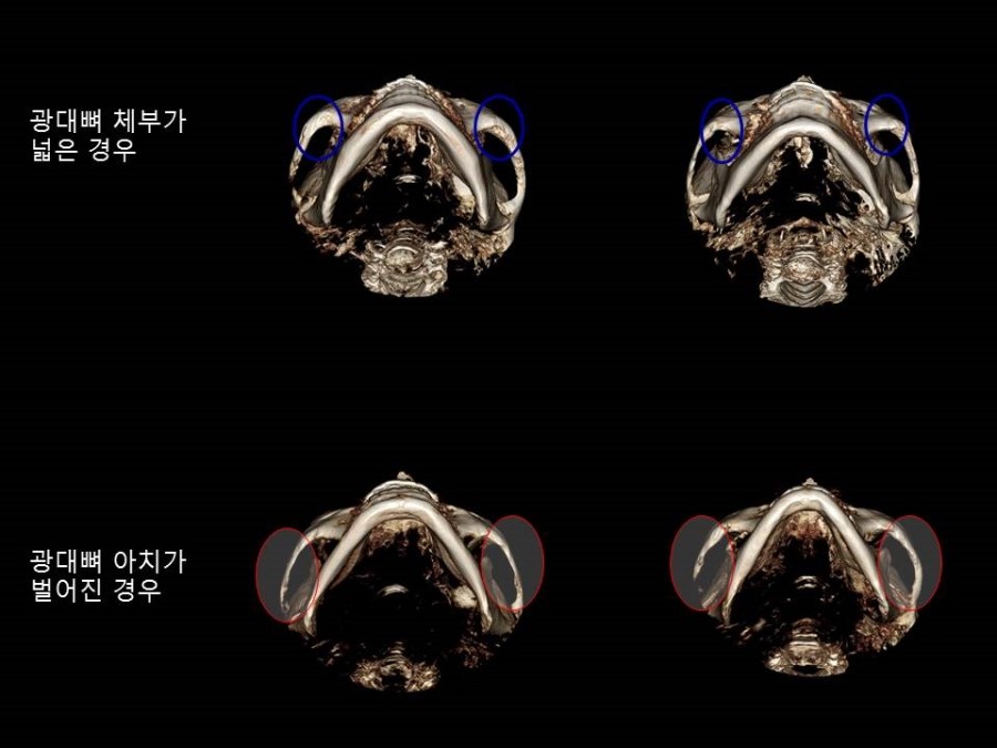
To analyze this posterior portion of the zygomatic arch, commonly called the back cheekbone, an exact reference point is needed.
The most reliable area to use as a reference point for the posterior zygoma is the temporomandibular joint.
Let us enlarge the CT images a little so that the temporomandibular joint and the posterior portion of the zygomatic arch can be seen clearly in each image.
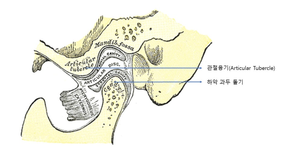
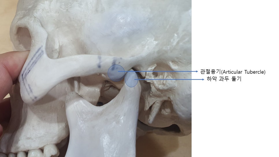
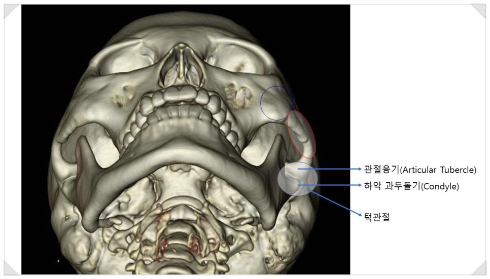
The temporomandibular joint is composed of the mandibular condyle, the temporal fossa, and the articular tubercle.
At this point, the most important thing to pay attention to in posterior cheekbone surgery is the need for a precise understanding of the position of the articular tubercle and avoiding structural damage to this area.
During the process of opening the mouth, the mandibular condyle rotates clockwise while also moving slightly outward, so if even part of the outside of the articular tubercle is removed, it may cause long-term temporomandibular joint instability.
Therefore, doctors who specialize in treating the temporomandibular joint place great importance on the articular tubercle.
If you have understood a little of the anatomical information about the temporomandibular joint area and the posterior zygoma, I will now explain effective posterior zygoma reduction.
For the posterior zygoma, including the 45-degree cheekbone and the side cheekbone, to be reduced effectively, it must be moved clearly farther inward than the mandibular condyle or the articular tubercle.
The reasons are as follows.
-
When considering the rotation of the zygomatic arch, in the arc rotated around the osteotomy site of the zygomatic body as the pivot point, the posterior part must move as much as possible for effective reduction to also be expected in the area near the pivot point.
-
The soft tissue behind the sideburns around the temporomandibular joint, such as the fat layer and muscles, is relatively thin, but the buccal fat covering the zygomatic arch is relatively thick. Therefore, a greater amount of bone movement is needed to expect a reduction effect that is actually reflected in the skin.
-
Visually, the position of the articular tubercle is inside the sideburns. Even if the area in front of the articular tubercle, which is hidden by the sideburns, is moved as much as possible, the visual prominence caused by the articular tubercle is hardly noticeable.
-
Of course, the shape of the zygomatic arch varies depending on the individual. However, in many cases, the portion just in front of the posterior cheekbone is more prominent than the posterior cheekbone itself. Therefore, the posterior cheekbone must be moved as far inward as possible to expect a reduction effect of 7 to 8 mm or more on one side of the face after surgery.
In this posterior cheekbone reduction surgery, if the posterior portion of the zygomatic arch is fixed at a position similar to that of the temporomandibular joint (the mandibular condyle), the actual reduction effect will be only about 3 to 4 mm.
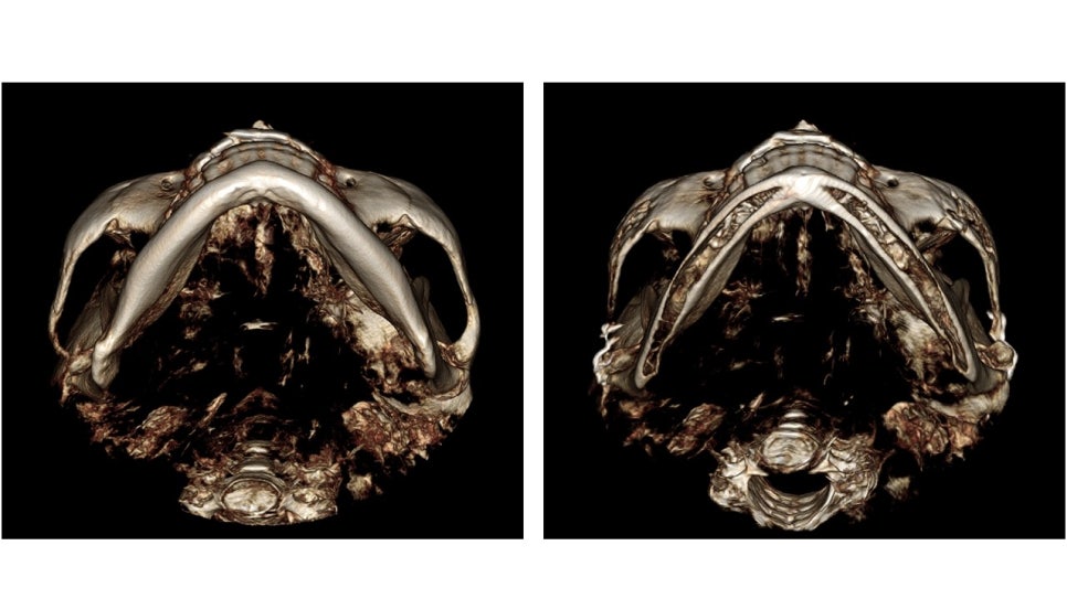
To compare the bone condition before and after surgery, it is necessary to check from various angles, and what is even more important is whether the conditions are the same -
it is important to check whether the thickness of the lower jawbone seen on the pre- and post-operative CT scans is the same, and whether the angle at which the arch formed by the lower jawbone opens is the same.
The most important parts of analyzing the posterior cheekbone reduction effect after zygoma surgery on CT are:
-
Set the position of the temporomandibular joint as the exact reference point
-
Check whether there is any damage to the articular tubercle
-
Check whether osteotomy has been performed in front of the articular tubercle and whether it has been moved farther inward than the temporomandibular joint
The more important point than these three is that it is above all important to confirm:
"from many different angles"
"under the same conditions"
"with accurate reference points"