In this post,
for those who are considering zygoma reduction surgery, or who have already undergone zygoma reduction surgery but are worried because the change in facial shape is different from what they expected,
I would like to explain the differences in postoperative facial changes depending on the method of zygoma reduction surgery.
Many people considering zygoma reduction surgery want to reduce the width of the face from the front, and I think they hope for a change toward a younger, more lively image while keeping a three-dimensional facial shape as the face becomes smaller.
"I still understand that many people think the methods of zygoma reduction surgery are more or less the same, and that the degree of change after surgery will also be similar.
However, the methods of zygoma reduction surgery are very diverse, and depending on the method, the direction of the change can be completely opposite."
Depending on the surgical method, people often complain that after surgery the face becomes a mantis-like face and they feel depressed, or that the width of the face does not decrease when viewed from the front and only the front cheekbone sinks in, making the face look older and saggy.
Let’s take a look through diagrams at what a mantis-type face or pyramid-type deformity related to zygoma surgery actually means.
This is also an expression I have heard from people who came in for facial contouring consultations in the past, but what exactly does a mantis-type face refer to?
As you can see in the figure below, a mantis-type face seems to refer to an inverted-triangle face shape.

Also, a pyramid-type face,
as shown in the diagram on the screen,
refers to the contour of the middle part of the face that becomes wider from the front toward the back when viewed from the front.
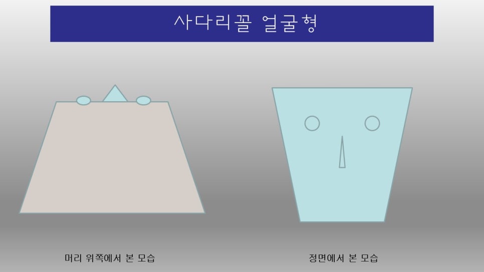
In reality, it would mean a face shape that becomes gradually wider when the face is tilted forward like this.
Then, let’s look at what direction zygoma reduction surgery should take in order to improve these issues, through a 3D simulation.
In the screen below, the left side shows a surgical method in which the front portion is partially cut and the far posterior part of the zygomatic arch is completely osteotomized and then rotated.
The right side shows a method in which part of the anterior bone is completely removed, the arch is osteotomized, and then the front and back are fixed with double fixation.
I think you will also be able to easily understand the difference between the two surgical methods and the difference in the bone appearance after surgery.
https://youtu.be/fr-ibTGj13g?si=i5dT2j_bl3qf_5sq
In this post, I briefly explained the differences in postoperative changes depending on the zygoma surgery method.
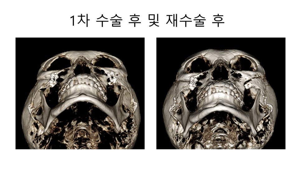
Lastly,
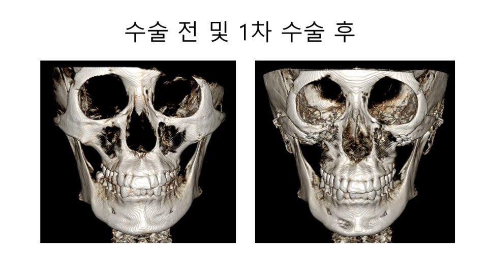
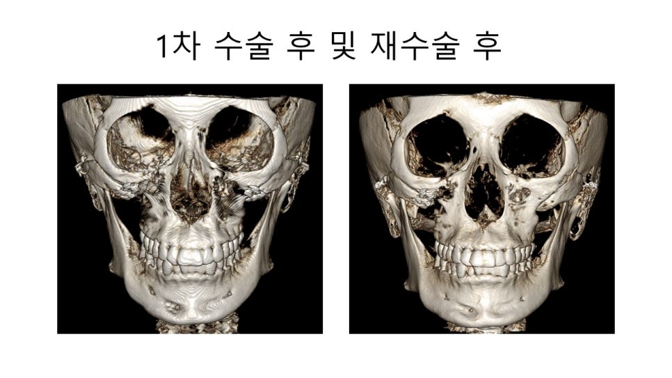
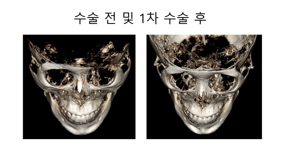
I am attaching preoperative and postoperative CT scans of a patient who visited me for a consultation several years ago, then underwent zygoma reduction surgery at another clinic, and recently came to me for a consultation regarding revision zygoma surgery.
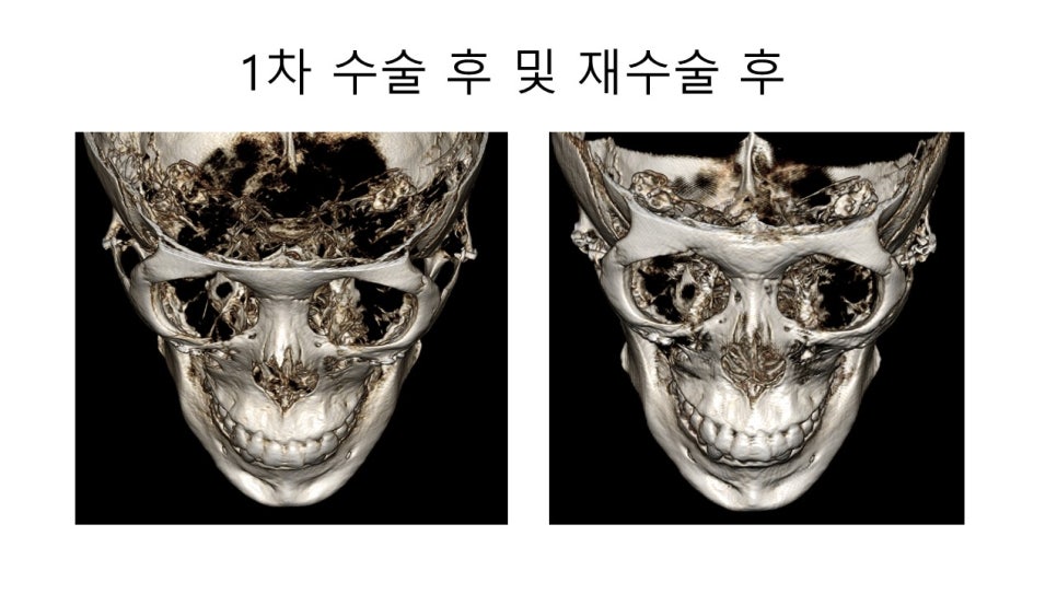
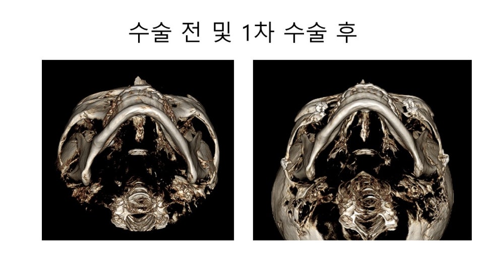
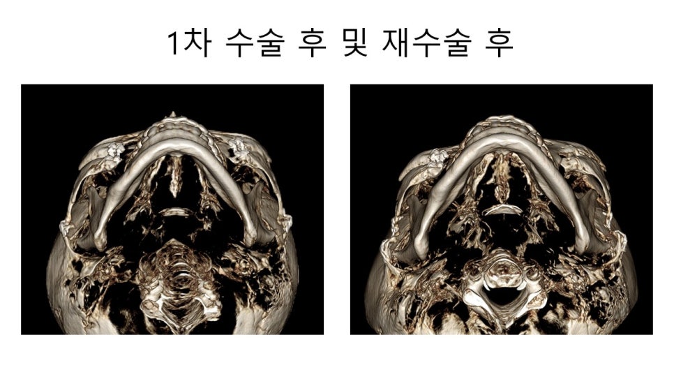
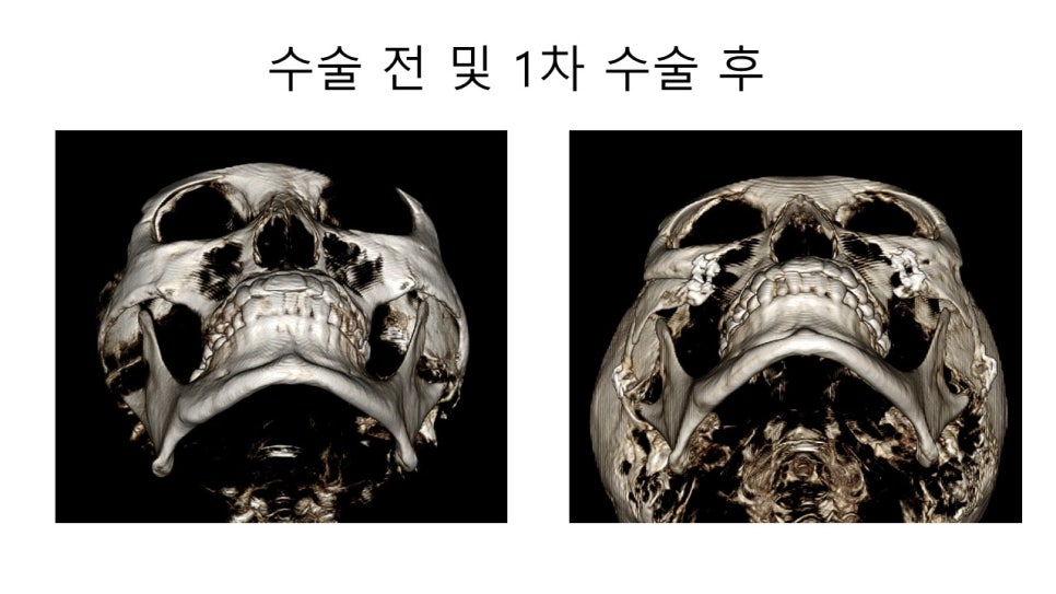
I hope this post will be helpful for those who are considering zygoma reduction surgery to improve the contour of the middle part of the face, or for those who are worried because the changes after zygoma reduction surgery turned out differently from the direction they had expected.