In the clinic, while meeting and consulting with many people who come in with various concerns about facial contouring surgery, one topic I have been hearing more often lately is the 3DCT images taken before and after surgery.
The level of knowledge some people have about facial contouring surgery is so specialized that I am sometimes surprised as well.
Many people have shown interest in the 3DCT images before and after my surgeries, so with the consent of the patients who received surgery, I have decided to post the 3DCT images of the patients I operated on little by little.
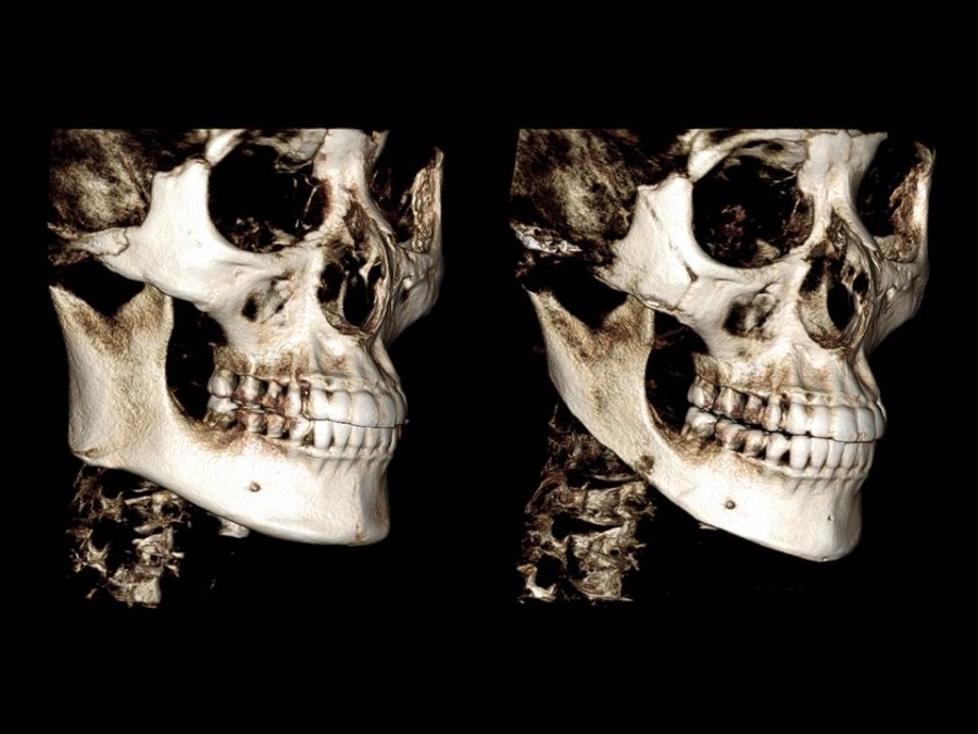
This is the change in the bone structure seen on the scan taken before surgery and two weeks after surgery for a patient I performed V-line square jaw surgery and zygoma reduction surgery on one month ago.
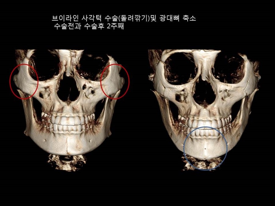
The outline of the widened cheekbone inside the red circle before surgery has moved inward after surgery, and on the 3DCT taken two weeks after surgery, only slight traces of the osteotomy line in the anterior zygomatic body are visible, indicating that continuity has been well maintained.
To make the front chin area smaller and more refined, I adjusted both sides evenly in the form of rotational shaving.
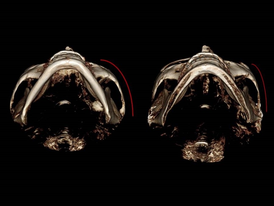
On the 3DCT taken from the base, the outline of the cheekbone arch, which had been widely spread before surgery, has moved inward after surgery.
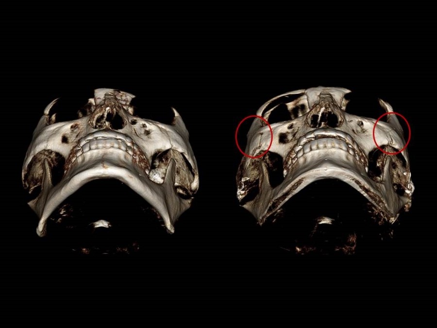
The part I consider most important in zygoma reduction surgery is maintaining the overall continuity of the cheekbone.
On the postoperative 3DCT, you can observe that continuity is maintained at the anterior osteotomy line inside the red line.