The facial outline appeared somewhat wide and angular, and the chin point was positioned toward the back. This is the 3D CT scan of a patient who had surgery with me one month ago, showing the preoperative state and the condition two weeks after surgery.
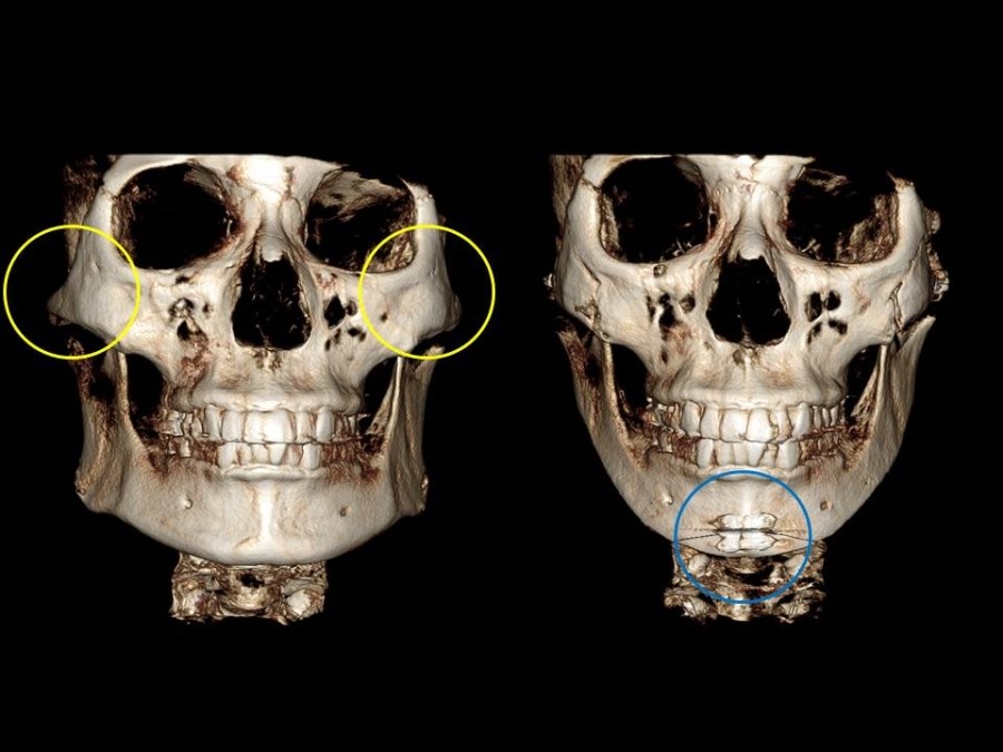
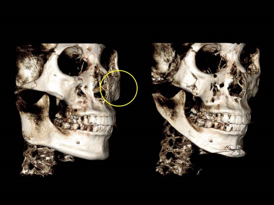
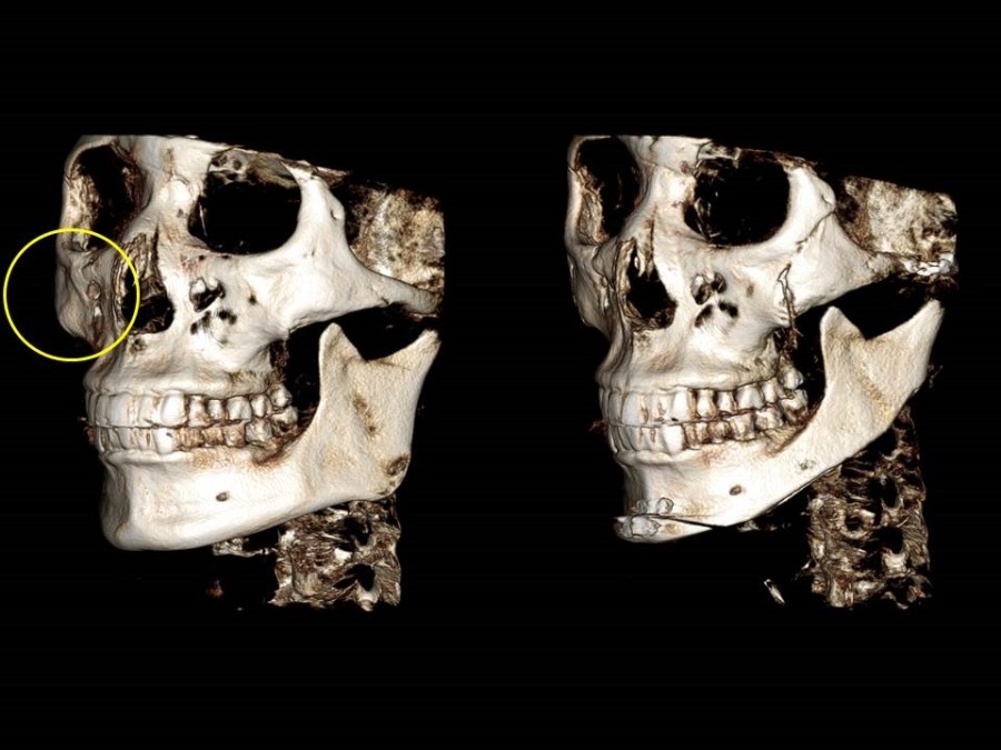
As you can see in the left image, the contour of the prominent cheekbones that were wide and flared before surgery (inside the yellow circle) has been brought significantly inward after surgery, based on the anterior osteotomy line. Since the right lateral zygoma was wider before surgery, I moved the right zygomatic arch inward more.
To make the angular lower jaw bone contour smaller and smoother, I reduced a large amount of the cortical bone and the body of the mandible, including the angular part of the lower jaw.
The front chin area was horizontally osteotomized and advanced forward, and it was carefully refined so that no step-off deformity would occur in the surrounding area.
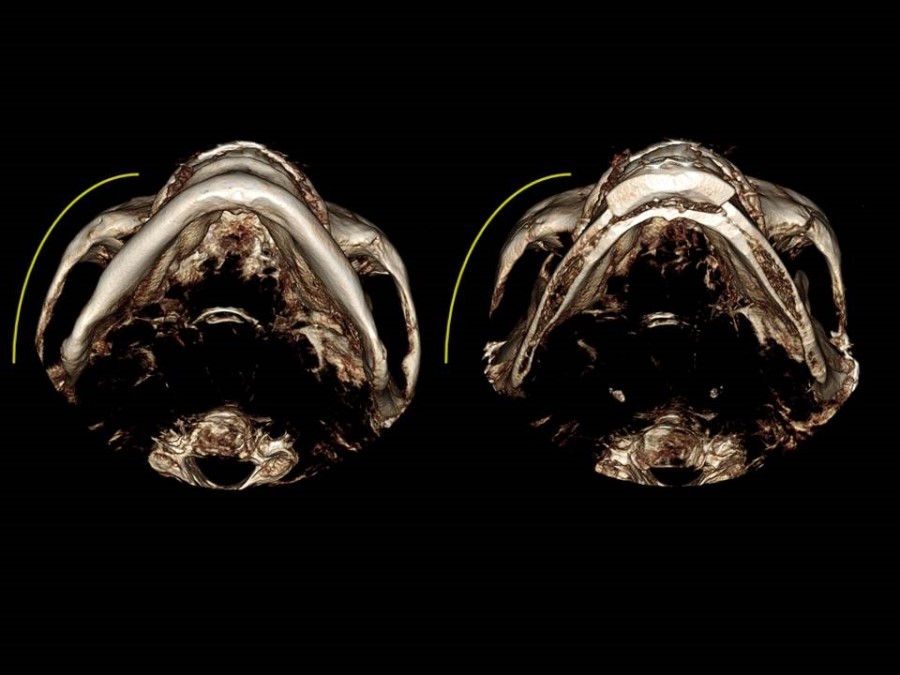
This image is viewed from below the chin area looking upward.
You can observe that the zygomatic arch, which was flared before surgery, has moved inward after surgery.

This is the angle that lets us check the continuity of the cheekbones, which I always consider the most important part of zygoma reduction surgery.
After surgery, based on the anterior osteotomy line, you can see that there is no step-off deformity in the area where the cheekbone body transitions to the zygomatic arch, and continuity has been maintained.