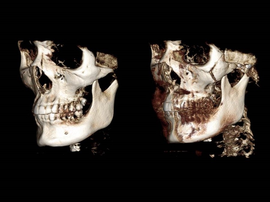
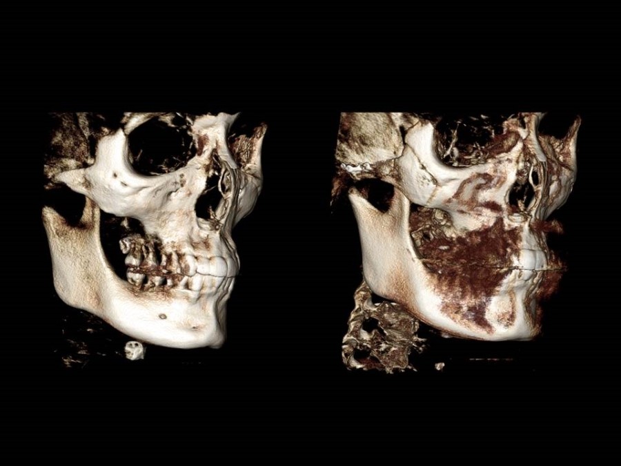
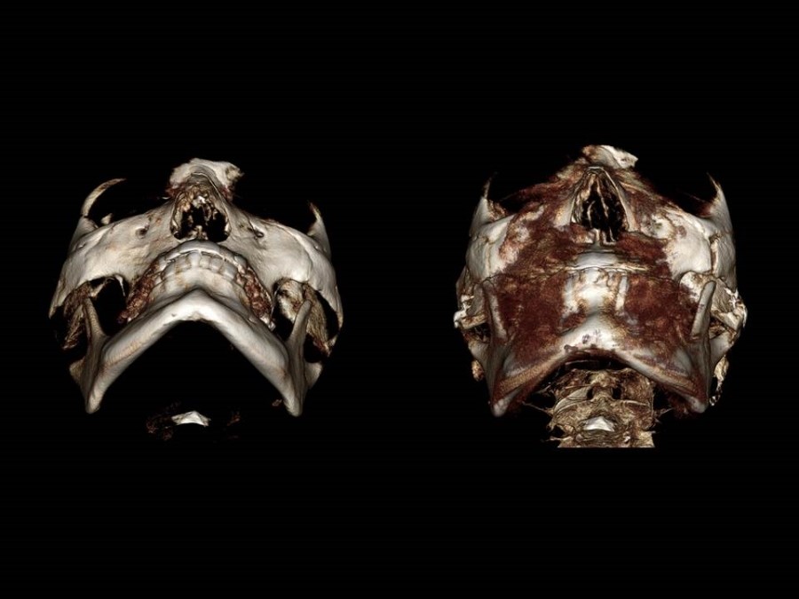
These are the pre- and post-operative 3D CT scans of a person who came from far away and underwent zygomatic reduction surgery on December 12, 2013.
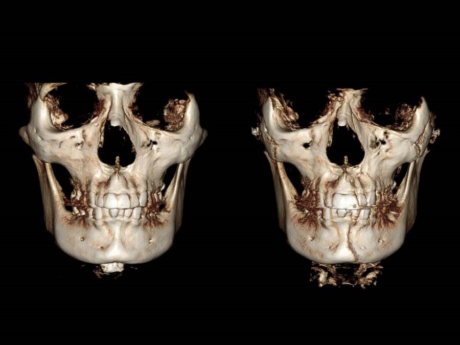
The left side is the pre-operative 3D CT, and the right side is the 3D CT taken two weeks after surgery.
The partial osteotomy line of the anterior zygomatic body appears to have fused almost completely even two weeks after surgery. In addition, the amount of movement of the lateral zygomatic bone after surgery shows that, based on the bone at the outer corner of the eye, the contour of the lower part of the zygomatic bone (the zygomatic body and arch region) has moved inward.
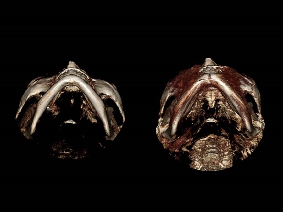
The left side shows the change in the contour of the zygomatic arch as seen from the base view (from below the chin looking upward) before surgery.
You can see that the position of the zygomatic arch, which looked widened before surgery, has moved inward after surgery, and it can be observed that continuity is maintained in the area where the front osteotomy line transitions to the zygomatic arch region.
This is the part I consider most important: it shows the continuity of the bone at the outer corner of the eye after zygomatic reduction surgery.
If a step-off occurs at the outer corner of the eye after surgery, as the swelling goes down and time passes, it may appear shadowed or sunken. In such cases, correction may not be possible even with methods such as fat grafting.