When reduction malarplasty is performed through a scalp incision, it is difficult to expect a significant reduction effect on the lateral cheekbones.
Therefore, in recent years, there have been more cases of people who previously underwent reduction malarplasty through a scalp incision considering revision surgery for lateral cheekbone reduction.
In such cases, I will explain the positional changes of the lateral cheekbones through preoperative and postoperative 3D CT analysis.
The cheekbone revision surgery was performed through an intraoral incision and an incision in the sideburn area. It was said that because there was little postoperative swelling, the reduction in the lateral cheekbones could be directly noticed two weeks after surgery, and there was no cheek sagging at all.
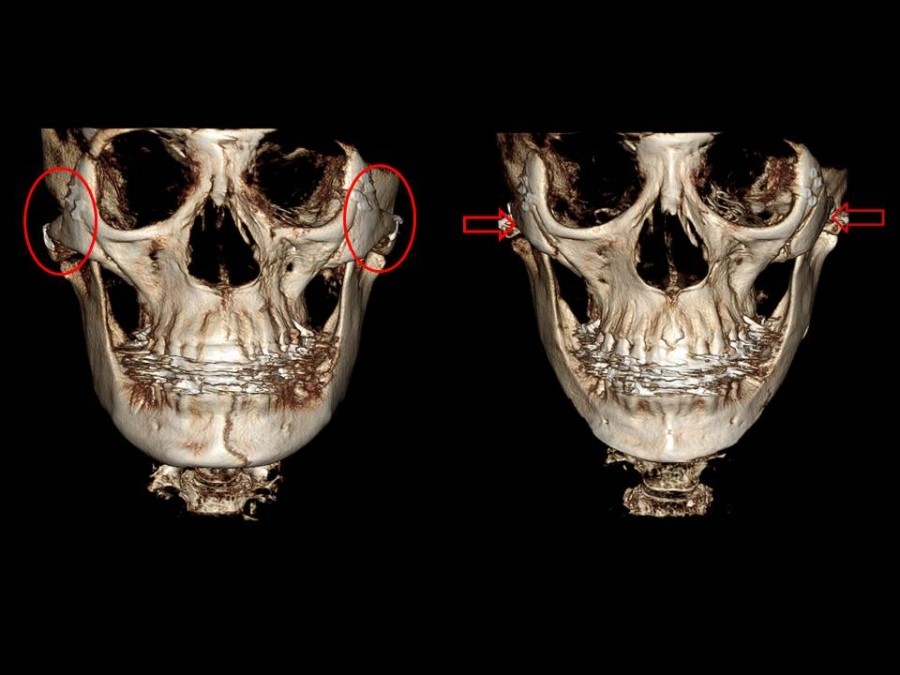
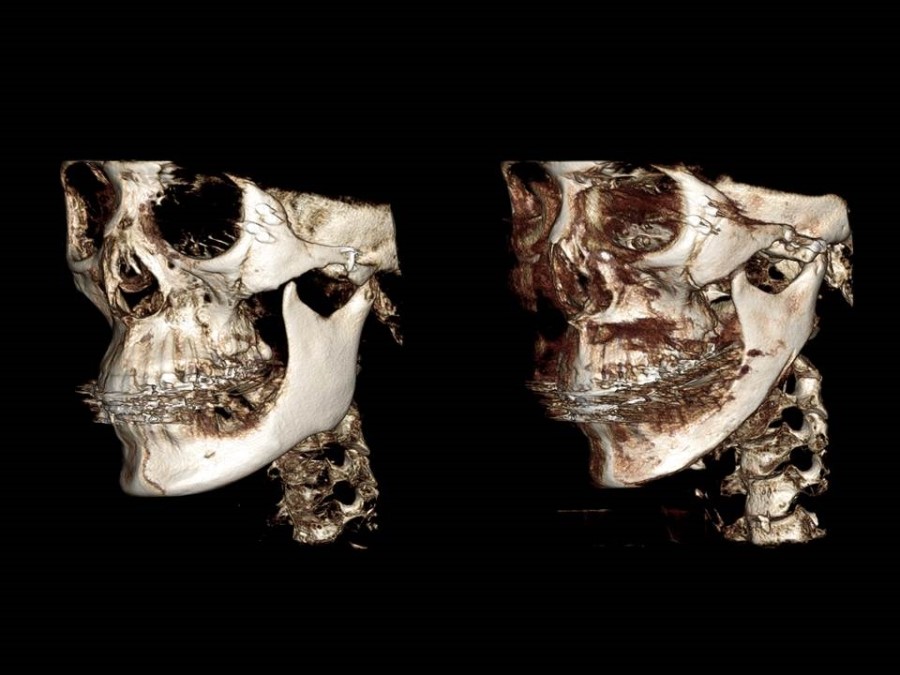
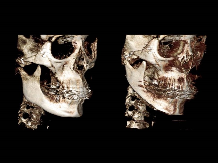
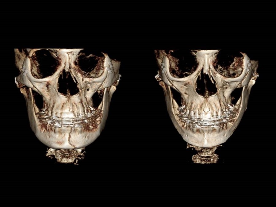
In the 3D CT above, the image on the left shows the appearance of the cheekbones after reduction malarplasty performed through a scalp incision, and the image on the right shows the result after partial osteotomy of the anterior body and osteotomy and fixation of the most posterior part of the zygomatic arch.
After surgery, it can be seen that the cheekbones below the outer corners of the eyes have moved inward, reducing the width of the face.

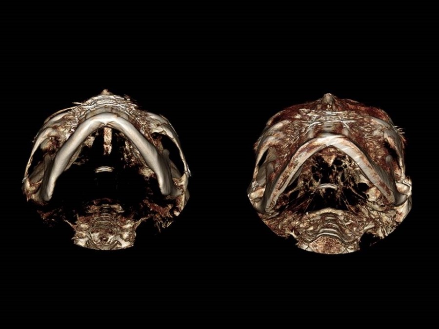
These images show the positional change of the zygomatic arch as seen from the base.
The zygomatic arch, which was spread outward before surgery, has been moved inward and fixed after surgery.