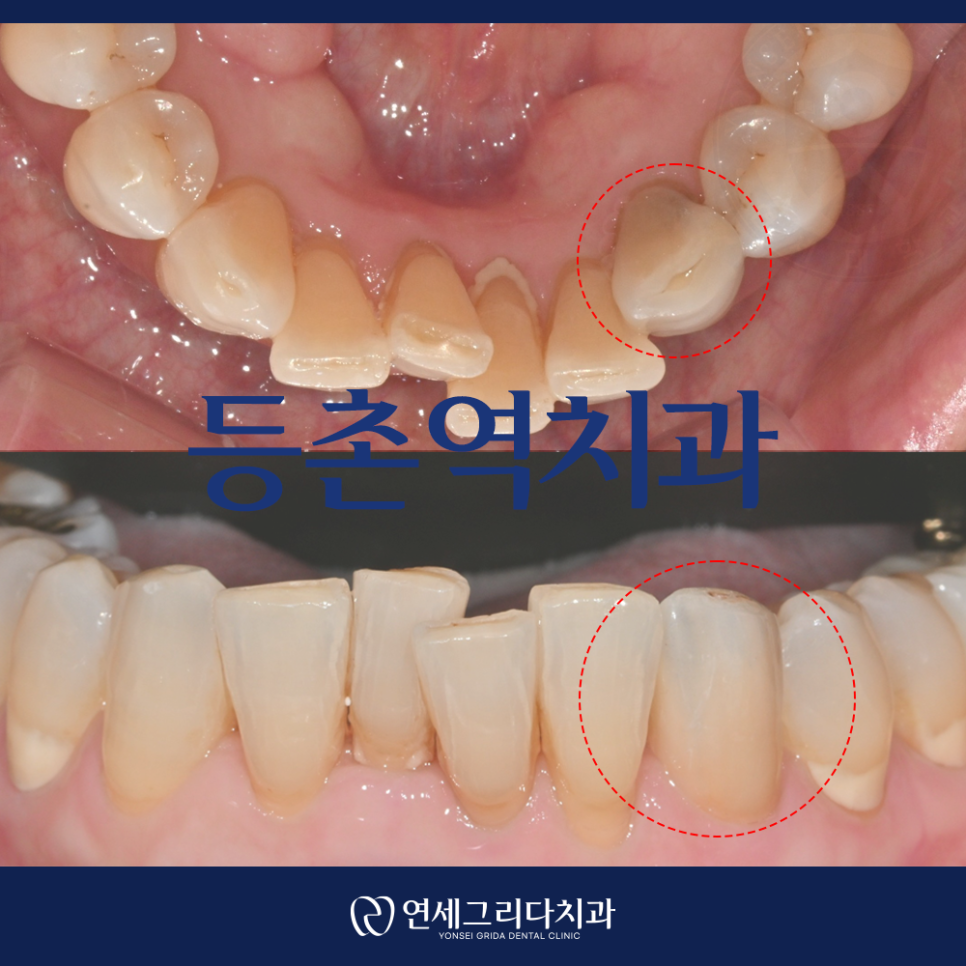
To keep your teeth healthy,
even if you do not have any teeth that feel uncomfortable at the moment,
it is important to visit the dentist regularly
to check your oral condition.
Many people think that there is no problem if their teeth do not hurt.
However,
because there are no nerves in the enamel
that surrounds the outside of the teeth,
you may often not experience any obvious symptoms
even when tooth decay is progressing.
If your teeth feel sensitive
or painful when you eat cold or hot foods,
the decay may already have progressed deep inside the tooth
and may have reached the area near the nerve tissue.
Even if no particular abnormality is visible to the naked eye,
it is important to have the condition properly checked
through a dental examination.
Because teeth can develop new problems due to factors such as diet, brushing habits, and gum condition,
regular checkups and consistent X-ray examinations
can be helpful.
Today, I would like to introduce the case of a patient
who visited us to have their condition properly checked
after being told during a health examination at another medical institution
that severe tooth decay had been found.
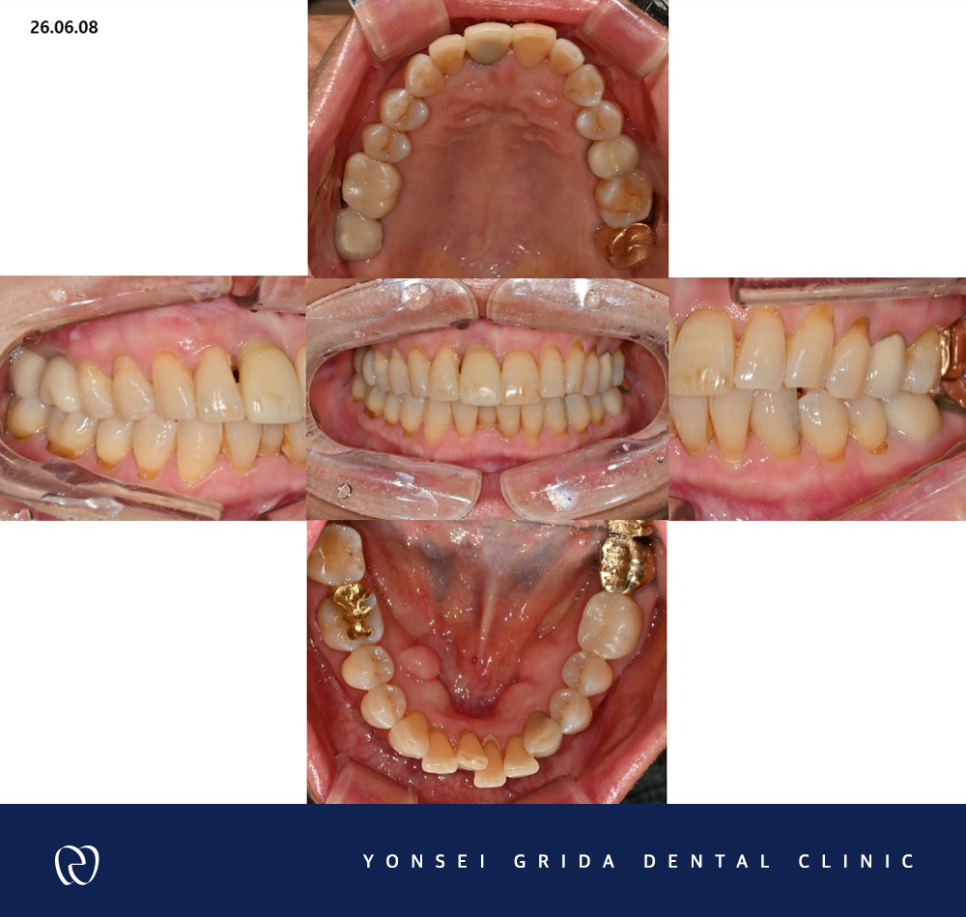
No major problem was visible to the naked eye
The patient who visited Yonsei Grida Dental Clinic
had been told at another dental clinic they usually visited
just three days earlier
that there was no particular problem with their dental condition.
However, they were quite surprised
after suddenly being informed during a health examination
that tooth decay was suspected.
The patient themselves had never experienced pain in daily life
or discomfort while eating,
so they wanted to clearly confirm
whether there was actually a problem with their teeth.
Accordingly, at the Deungchon Station dental clinic,
we decided to conduct examinations including radiography
to carefully assess the patient's oral condition
and check even areas inside the teeth
that could not be seen with the naked eye.
The images revealed an area requiring further examination
around the lower canine,
so we performed X-rays
to assess the condition of that area more accurately.
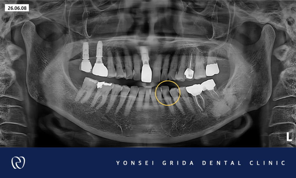
Full panoramic X-ray image
The X-ray examination confirmed
that extensive tooth decay was progressing
in the lower left canine.
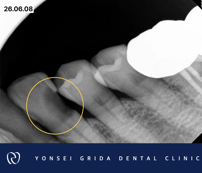
Although no major abnormality was found
when the tooth was examined with the naked eye,
the decay had progressed considerably deep inside the tooth
and had affected the pulp,
the nerve tissue in the center of the tooth.
Therefore, once all the decayed and infected areas were removed,
we expected that not much healthy tooth structure would remain
to support the tooth.
When a substantial portion of the crown is damaged in this way,
simply filling the empty space
may make it difficult for the tooth to withstand
the forces transmitted during chewing.
Therefore, it was necessary to establish a step-by-step treatment plan
to protect the remaining tooth
and ensure that the prosthetic restoration placed afterward
could remain stable.
✔ First, we decided to perform root canal treatment
to remove the infected nerve tissue
inside the lower left canine
and clean and disinfect the root canal.
✔ After the root canal treatment was completed,
a post would be inserted
to reinforce the empty interior of the tooth
and support the upper structure.
✔ Next, core material would be used
to rebuild the damaged crown
and create a foundation
that could support the prosthetic restoration.
✔ In the final step,
a crown prosthesis would be placed
to cover and protect the entire reconstructed tooth
and restore its chewing function.
The treatment process described above
was a comprehensive decision intended to preserve
as much of the remaining natural tooth as possible
and allow the patient to use it stably even after treatment.
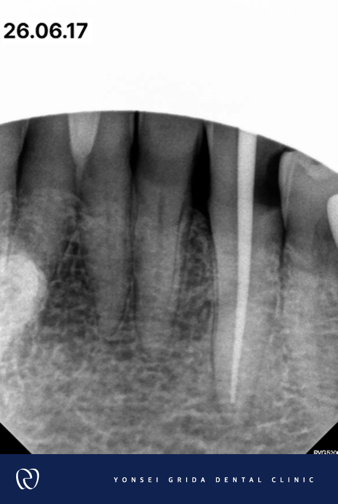
During the root canal treatment performed in stages as described above,
we carefully controlled any stimulation or pain
that the patient might experience
and safely removed the infected tissue.
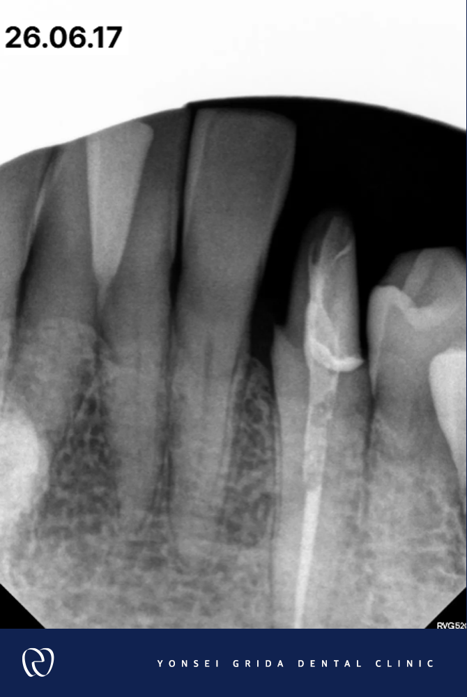
After placing a post to support the weakened tooth,
we firmly filled the central area
and completed a shape that could receive the prosthetic restoration.
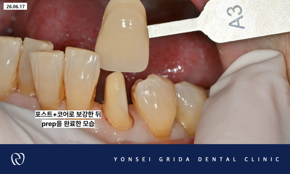
When fabricating the final crown,
we focused on creating a shape and color
similar to those of a natural tooth,
taking harmony with the surrounding teeth into consideration.
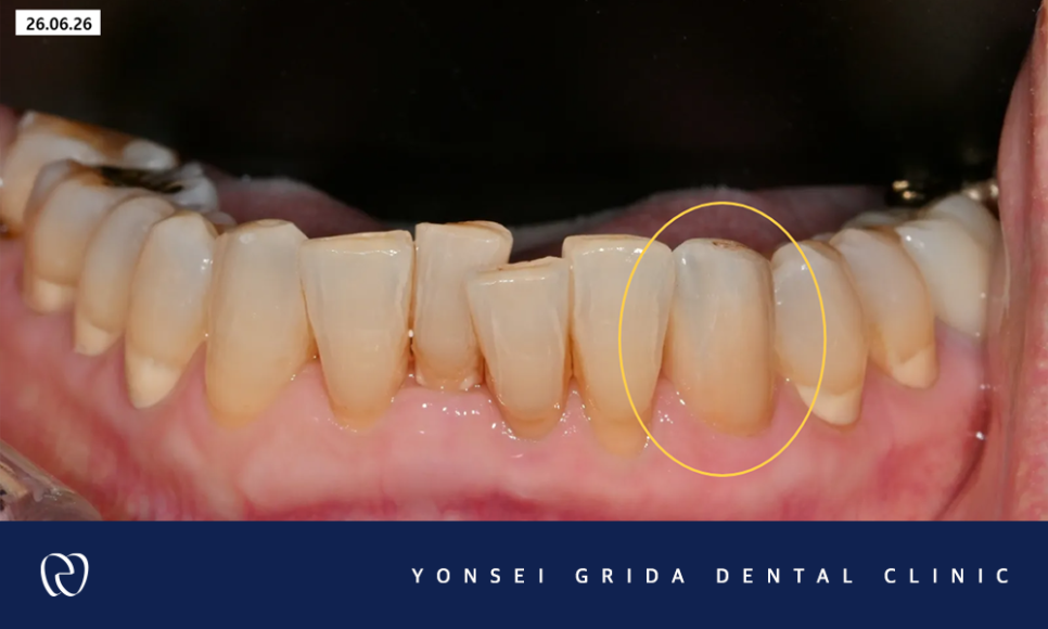
This treatment
was completed on June 26, 2026.
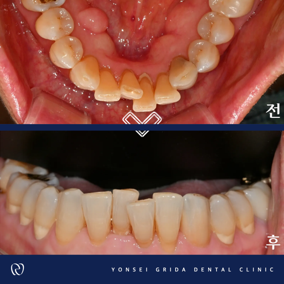
The patient said that they received treatment comfortably
without experiencing pain or discomfort during the process
and expressed great satisfaction.
In particular, they expressed their deep appreciation
for the fact that the final crown prosthesis
looked so natural and aesthetically pleasing
that it was difficult to distinguish from the original natural tooth.
The case introduced today by the Deungchon Station dental clinic
confirmed the importance of checking for problems inside the teeth
in advance through an examination,
even when no particular pain or discomfort is felt.
Even if the teeth appear normal to the naked eye,
a condition may be progressing inside the tooth.
Checking the condition at the appropriate time
and beginning treatment suited to the oral situation
may help preserve the natural tooth.
Yonsei Grida Dental Clinic
carefully examines each patient's different oral condition,
provides a thorough explanation of the treatment currently needed,
and provides care accordingly.
We do not take even small changes lightly.
From the patient's perspective, we take another careful look
and thoroughly examine areas that are difficult to see immediately,
striving to guide each patient toward an appropriate treatment direction.
The Deungchon Station dental clinic
will continue to improve its clinical capabilities
to protect patients' oral health over the long term
and repay their trust
with warm and attentive care.
Thank you.
*All case photographs were posted with the consent of the individuals concerned, in accordance with Articles 23 and 56 of the Medical Service Act.
*All treatment photographs are of patients treated at this clinic.
*All treatment photographs are of the same individual and were taken under the same conditions.
*Depending on individual differences, side effects may occur after procedures or surgery. Please consult thoroughly with your medical professionals.