Pneumothorax is a term formed by combining the Chinese character for “air,” “gi” (氣), and the character for “chest,” “heung” (胸).
Pneumothorax refers to a condition in which air enters the pleural cavity (thoracic cavity) surrounding the lungs for various reasons, causing symptoms such as shortness of breath and chest
pain.
- Anatomical structure and function of the respiratory system
To understand the disease called pneumothorax, you need to have a good understanding of the structure of the respiratory system and the principles of breathing movements. The human
respiratory system is composed of the organs and tissues needed for breathing, and their names and roles are as follows.
- Airway
The airway serves as a passage that allows oxygen-rich outside air to enter the lungs and expels carbon dioxide produced inside the body to the outside. The airway consists of the nasal cavity connected to the nose, the oral cavity connected to the mouth, the larynx, the trachea, and the bronchi.
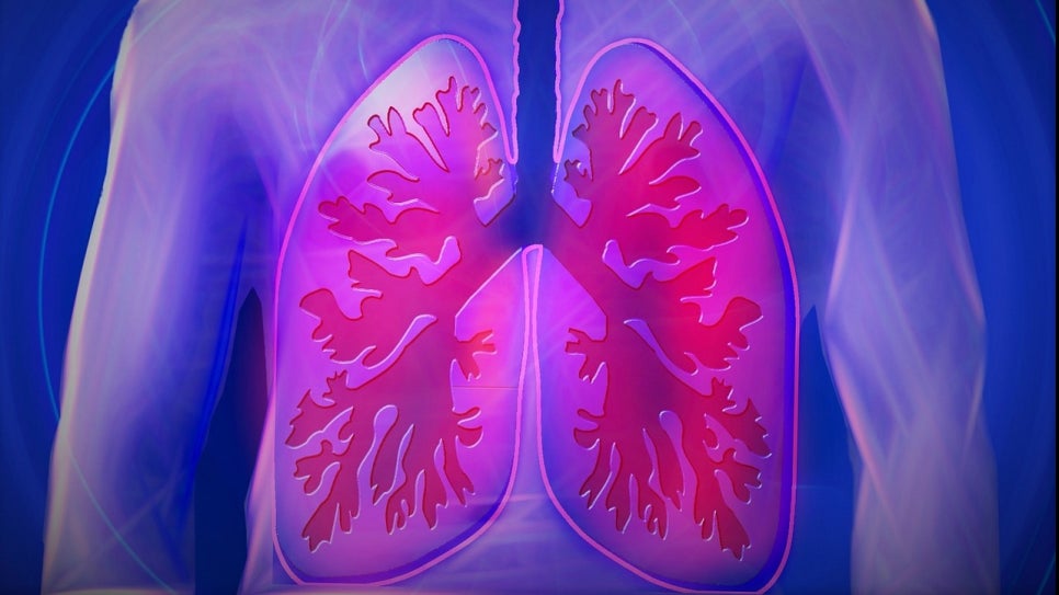
- Lungs and blood vessels
Inside the lungs, the bronchi, which make up the ends of the airway, branch out and become progressively thinner to form the bronchioles, and
these lead to the alveoli, hollow sac-like structures resembling a bunch of grapes. Around the alveoli, capillaries
surround them like a net, where gas exchange takes place: oxygen entering through the bronchi is absorbed, and carbon dioxide produced in the body is
released outside the body.
- Thoracic cage and respiratory muscles
The box-shaped space that surrounds and protects the lungs is called the “thoracic cage” (胸廓). The thoracic cage is surrounded by the spinal bones at the back, the sternum (breastbone) at the front, and the ribs on the sides. Meanwhile, the lower part of the thoracic cage is closed off by a muscle called the diaphragm, which separates the chest and abdomen. Between each rib, there are muscles called the intercostal muscles, and the contraction and relaxation of these respiratory muscles produce the breathing movements by which a person inhales and exhales.
- Pleura and pleural cavity (thoracic cavity)
Both lungs and the thoracic cage are surrounded by a thin membrane called the “pleura.” The pleura that covers the chest wall is called the “parietal pleura,” and the pleura
that covers the lungs is called the “visceral pleura.” The empty space between the pleura is called the “pleural cavity (thoracic cavity) (腔),” and
in a healthy person, only about 10–15 cc of pleural fluid (胸水) is present inside the pleural cavity (thoracic cavity).
- The concept of pneumothorax
When air accumulates in the pleural cavity (thoracic cavity) for some reason, the lungs are compressed and collapse, making it impossible to perform proper
breathing movements. This is called “pneumothorax.”
Normal state
· When the thoracic cage expands due to the diaphragm and intercostal muscles, outside air enters the lungs; conversely, when the thoracic cage contracts,
a breathing movement occurs in which air inside the lungs is expelled to the outside.
Pneumothorax state
· If the chest wall is damaged and the atmosphere communicates with the pleural cavity (thoracic cavity), outside air enters the pleural cavity (thoracic cavity) through the parietal pleura, or if the visceral pleura is damaged and air from the alveoli leaks into the pleural cavity (thoracic cavity), a pneumothorax state occurs.
The lungs collapse, and even if the thoracic cage expands or contracts, proper breathing movements cannot occur, leading to shortness of breath.
So far, we have explained pneumothorax.
In the next part, we will look at the causes and risk factors of pneumothorax.
Source: Korea Disease Control and Prevention Agency National Health Information Portal