Because different parts of the brain are responsible for different functions, injury to a specific area can cause characteristic neurological symptoms. Doctors check the overall condition, such as blood pressure, and the state of consciousness, and use various neurological tests to determine whether a stroke has occurred, the extent of the damage, and the location of the damage. In recent years, various tests have been developed that can diagnose stroke and accurately assess the location and extent of damage, and they are being actively used in patient diagnosis and treatment.
- Computed Tomography (CT)
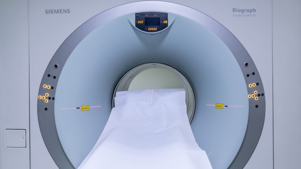
CT is the most commonly used test for diagnosing stroke. It uses X-rays to image the body and a computer to create cross-sectional images of the body.
The advantage of this test is that it can be performed relatively quickly and can rapidly distinguish whether there is a brain hemorrhage. However, in some cases a brain tumor may look like a brain hemorrhage, and in ischemic stroke, the lesion may not appear until a certain amount of time has passed after onset, which can make diagnosis difficult. In hemorrhagic stroke, bleeding can be observed on CT immediately after it occurs, so it is useful as a tool for ruling out brain hemorrhage before using thrombolytic drugs to treat ischemic stroke. In addition, CT scans are also important for monitoring the course of brain hemorrhage, which can occur as a complication even after thrombolytic drugs are used.
- Magnetic Resonance Imaging (MRI)
Magnetic resonance imaging (MRI) is a test widely used along with CT for stroke evaluation, and it obtains cross-sectional images of the body using magnetic fields. Compared with CT, MRI has similar ability to diagnose bleeding, but it is much more useful for diagnosing early ischemic cerebral infarction, small infarctions, and brain tumors that may resemble brain hemorrhage. However, patients with metal attachments such as pacemakers cannot undergo the test, and it is relatively more expensive than CT. Meanwhile, MR angiography, which uses MRI to image the state of blood vessels, can easily check the condition of the brain's blood vessels.
- Angiography
Angiography is a test that obtains images of blood vessels by injecting a contrast agent that does not allow X-rays to pass through the blood vessels while taking X-ray images. Since stroke is a disease caused by blocked or ruptured blood vessels, directly checking the condition of blood vessels—such as where they have narrowed or become blocked—through images such as angiography helps in diagnosis and treatment.
For angiography, a hollow thin catheter is first inserted through the carotid artery, subclavian vein, brachial artery, or femoral artery, and advanced to the starting point of the vessel to be imaged. Then, a small amount of contrast agent is injected through the catheter with a syringe while images are taken. At this time, because the blood vessels through which the contrast agent is flowing do not allow X-rays to pass through as much as other tissues, the shape of the blood vessels appears distinguishable in the images.
Angiography has the advantage of showing blood vessels accurately, but it is an invasive test and, although uncommon, complications such as unexpected vessel occlusion can occur during the procedure. For this reason, noninvasive tests such as MR angiography are now widely used to examine cerebral blood vessels.
- Ultrasound and Heart Tests
Ultrasound is a method in which a device that generates ultrasound waves sends them into the body, and the waves reflected back from each tissue are assembled into an image.
For the diagnosis and evaluation of stroke, two tests are mainly used: carotid ultrasound and echocardiography.
- Carotid Ultrasound
Carotid ultrasound is a test that uses an ultrasound diagnostic device to measure the condition of the carotid arteries that supply blood to the brain. Carotid ultrasound can check whether a thrombus has formed inside the vessel, the degree of narrowing of the vessel, and the speed of blood flow through the carotid artery.
- Echocardiography and Arrhythmia Testing
Echocardiography is a method of directly observing the inside of the heart moving in real time using an ultrasound diagnostic device, checking the heart's structure and evaluating its hemodynamic function.
The most important purpose of echocardiography is to check for the presence of thrombi inside the heart. This is because in patients with abnormalities in heart function, such as atrial fibrillation, blood flow inside the heart may become stagnant and thrombi may form, and if part of a thrombus breaks off and travels through the arteries to block a cerebral blood vessel, it can cause an ischemic stroke.
In addition, echocardiography is also used to diagnose congenital abnormalities of the heart and great vessels, cardiac enlargement, myocardial hypertrophy, abnormalities in heart muscle movement, the presence and severity of valvular disease, and abnormal structures inside and around the heart.
In addition to echocardiography, electrocardiography may also be performed for 24 hours or longer to check for cardiac arrhythmias. If a cardiac arrhythmia such as atrial fibrillation is present, the risk of stroke is considered to increase more than fivefold, so active medication treatment is necessary.
So far, I have explained the diagnostic methods for stroke.
In the next part, we will look at stroke treatment.
Source: Korea Disease Control and Prevention Agency, National Health Information Portal