Symptoms
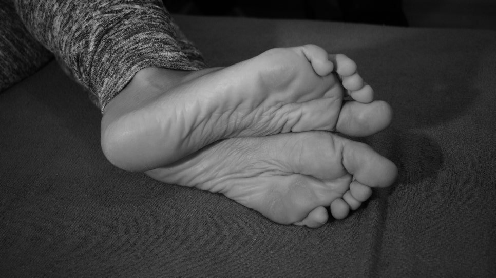
In most cases, there are no symptoms. When pain does occur, it is vague and dull, and pain may develop in the foot, ankle joint, and lower limbs, leading to a feeling of chronic fatigue. In children, they may avoid running or walking long distances, and dislike physical activities. In adults, the medial arch of the foot disappears visually, and the heel tilts outward. The inside of the shoe tends to wear out more, and pain may be felt during prolonged walking and exercise.
In children with flatfoot, deformation rather than foot pain is the main symptom, and pain is usually absent, but some may complain of pain after adolescence. Flexible flatfoot in children usually does not cause problems even if left untreated. In addition, patients who had pain generally become symptom-free as they grow.
Pain appears in relation to muscle fatigue after prolonged walking and exercise, excessive stretching of the plantar fascia, and so on. However, in cases of rigid flatfoot, pain may occur due to the underlying disease, or the foot may sprain frequently. In particular, pain and foot dysfunction related to posterior tibial tendon dysfunction may also occur.
Diagnosis
Diagnosis is made through a medical history review and physical examination of the visible deformity, along with plain radiography.
Let’s look at the diagnosis according to each cause of flatfoot.
- Medical history and physical examination
- Flexible flatfoot
Check whether the foot tires easily or is painful. When flatfoot is present under weight-bearing, if the arch appears when the big toe is lifted upward, it is called flexible; if it does not appear, it is called rigid.
As a general flexibility test, examine hyperextension of the elbow joint and knee joint, hyperextension of the metatarsophalangeal joints, and whether the thumb can touch the forearm. Also, to determine whether the Achilles tendon is shortened, measure the range of dorsiflexion of the ankle joint with the knee joint flexed and extended.
- Dysfunction of the posterior tibial tendon
It usually occurs in one foot and progresses chronically and gradually. The function of the posterior tibial tendon is evaluated through a heel rise test. If there is severe functional loss, the person cannot raise the heel. The posterior tibial tendon causes the heel to turn inward as it is lifted, and this phenomenon is observed.
- Congenital tarsal coalition
It is also called rigid flatfoot, but not all tarsal coalitions cause flatfoot. Rheumatoid arthritis can also cause rigid flatfoot, and traumatic flatfoot is also mostly rigid flatfoot. Symptoms appear when the joint is connected by bone, restricting movement, or when there is slight movement due to fibrous or cartilaginous union. Movement limitation mainly causes difficulty walking on uneven surfaces and pain while walking. Tarsal coalition includes calcaneonavicular coalition between the calcaneus and navicular bone, talocalcaneal coalition between the talus and calcaneus, and naviculocuneiform coalition between the navicular and cuneiform bones. Bone scan may also be helpful for diagnosis.
- Plain radiography
All measurements are taken in a standing position with weight-bearing.
- Anteroposterior view
(1) Talonavicular coverage angle
(2) Talar-first metatarsal angle
(3) Talocalcaneal angle
- Lateral view
(1) Talar-first metatarsal angle
(2) Talocalcaneal angle
(3) Talar horizontal angle
(4) Calcaneal pitch angle
- Congenital tarsal coalition
This is a condition in which two or more tarsal bones in the foot are congenitally connected to each other, and it is one of the most common causes of peroneal spastic flatfoot. Even when tarsal coalition is present, there are usually no symptoms, and it is often found incidentally on plain radiographs. Symptoms usually appear after puberty, and the condition varies from cases with almost no deformity, such as the heel turning outward or the medial arch disappearing, to severe cases. This abnormal fusion can occur between any bones, but talocalcaneal coalition, in which the calcaneus and talus are fused, is reported most commonly. If the patient stands on the cassette, bends the knee, and the X-ray beam is directed into the heel at an angle of 30–40 degrees to the cassette, coalition of the middle joint can be seen.
If the fused area is not clearly visible on the axial view, secondary signs may be helpful. Secondary findings include: ① a beak-like osteophyte on the dorsal aspect of the talar head, ② widening and rounding of the lateral talar process, ③ narrowing of the posterior subtalar joint space on the lateral view, and ④ the C sign (on the lateral view, the bony shadow of the talar beak and the fused bone appears continuous, forming a semicircle). However, not all of these findings are always visible.
For the diagnosis of tarsal coalition, computed tomography (CT) is the most accurate, and magnetic resonance imaging (MRI) has the advantage of showing fibrous union before the tarsal bone has ossified.
So far, we have explained the symptoms and diagnosis of flatfoot.
In the next part, we will look at the treatment of flatfoot.
Source: National Health Information Portal, Korea Disease Control and Prevention Agency