Hello. I am Kang Dong-ho, director of Yonsei U-Line Dental Clinic, Sillim Branch, located near Exit 7 of Sillim Station in Sillim-dong, Gwanak-gu.
Patients often ask me:
> “Doctor, I had a gold crown placed a while ago. How can I tell whether the tooth underneath has decayed?”
>
> “I’ve heard of secondary cavities, but I’m not really sure what they are.”
That’s right. Many patients who have received crown treatment feel anxious because they cannot see the condition underneath with their own eyes.


In fact, metal crowns do not allow X-rays to pass through easily, so even with imaging, the area underneath cannot be seen clearly. So how can we determine whether a cavity is developing underneath?
Could it be decayed inside even if it looks fine outside?
A crown is, literally, a “lid” that covers the tooth from the outside. Although the exterior is firmly covered with metal or ceramic, the tooth underneath is still the patient’s natural tooth.
If the cement has loosened or a gap has formed along the margin, food particles or bacteria can enter and cause decay. This is called a “secondary cavity.”
The problem is that even when decay progresses underneath in this way, there may be no visible symptoms on the outside.
If there is no pain, looseness, or discomfort when chewing, it can be even easier to overlook.
So how does the dentist check for it?

The basic but important “explorer examination”
Cavities visible from the outside can usually be identified easily by looking at them or taking an X-ray. However, it is difficult to examine the area beneath a restoration such as a gold crown. This is when an explorer examination is used.
During an explorer examination, the dentist follows the margin where the crown meets the tooth and gently presses each area with an instrument.
A gap that can be felt along the margin, an instrument sinking deeply into one spot, or a surface that feels rough rather than smooth may be warning signs.

If no particular abnormality is found with the explorer and the X-ray shows no signs of inflammation or other problems in the surrounding bone, the dentist may conclude, “It looks fine for now.” Of course, this means that no problem is apparent—it does not provide 100% certainty.
Is it really impossible to know whether the area under a gold crown has decayed?
Many people wonder about this:
> “Can’t it be checked accurately? Couldn’t you just remove the crown and look underneath?”
That is possible. Removing the crown allows the dentist to examine the area underneath directly.
However, a crown that has been removed usually cannot be reused and must be newly made. Because of the way the cement works, it is almost impossible to remove a crown and then bond it back in place.
For that reason, deliberately removing a crown simply to check underneath is not recommended, either practically or economically.
This leads to an important principle: have regular examinations, and treat the tooth when there is reason for concern.
Small signs—such as an unusual sensation, swollen gums, or food repeatedly getting stuck—can actually be important indicators.

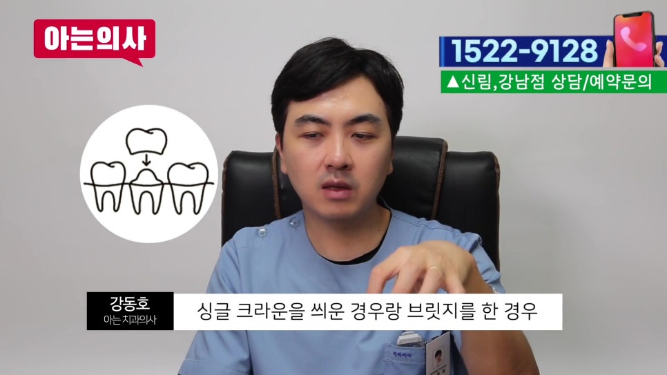
Bridges require even more care
When a bridge is involved, the situation is more complicated.
A bridge has no tooth in the middle and connects the teeth on both sides, which serve as supporting pillars.
When chewing forces are applied, this structure can move slightly, and the cement on one side may come loose in the process. The problem is that it can be difficult for the patient to notice this.
There may be no visible sign, and the explorer may not catch on, even though decay may already be progressing underneath.
In such cases, the dentist may gently hold the crown and move it slightly to check visually for subtle movement or to see whether one side has lifted.
However, because the movement can be extremely subtle, it is not easy to detect. When there is no clear evidence, the dentist may sometimes have no choice but to say, “Let’s monitor it for a while.”
That is why regular checkups are especially important for people with bridges. Decay does not necessarily cause pain right away.
As the decay progresses, symptoms such as an odor, looseness, or a foreign-body sensation may appear if the tooth still has a nerve. By then, the damage may already have progressed considerably.
That does not mean you need to replace it automatically every 10 years
This question also comes up:
> “Wouldn’t it be enough to replace it once every 10 years on a regular basis?”
In theory, that is possible. In reality, however, patients may naturally think, “Why replace something that still works?” From the dentist’s perspective, it can also easily be misunderstood as unnecessary treatment.
There is no reason to remove a restoration that is still functioning well.

That is why regular examinations are important.
If there is no abnormality, there is no need to intervene unnecessarily. If something appears suspicious, the dentist can assess it at that time and decide whether treatment is needed.


Visit the dentist regularly—small talk matters, too
In fact, it is not easy to visit the dentist even once a year.
But these small, routine visits are truly important.
> “It feels a little uncomfortable lately.”
>
> “It didn’t used to be like this, but food keeps getting stuck.”
Each of these small comments provides important information to the dentist.
The patient’s sensations, instincts, and changes in daily life—these details from the medical history can play a very important role in deciding whether treatment is needed.
Through regular checkups, the dentist can determine whether to say, “It looks fine for now, so let’s keep monitoring it,” or, “It is now time for treatment.”
Restorations, especially crowns and bridges, can change silently underneath.
They may look fine on the outside while the situation underneath is different.


So pay a little more attention to the condition of your teeth, and do not leave too much time between dental visits.
Please do not overlook warning signs that begin with small changes.