Depending on an individual’s eye shape, eye depth, distance between the eyebrows and eyes, and eyelid skin, there are suitable surgical methods. If surgery is performed with a method that does not match the person’s eye type, it can lead to an unnatural appearance or require revision surgery, so it is better to proceed with a suitable design and surgical method determined through detailed consultation.
Therefore, considering the individual’s eye type, current conditions, and facial harmony, the ideal angle, length, and degree of canthal opening that can create the best harmony should be carefully determined through consultation, and then the appropriate surgical method should be applied. In this post, we will talk about how the procedure was applied according to the eyes and facial types of different patients.
- A sharp eye shape
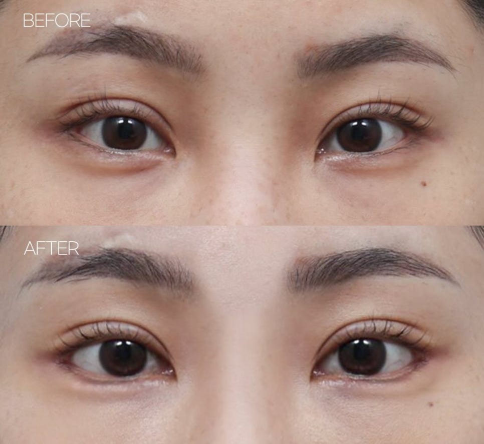
Eyes with a high inner-to-outer corner angle can give a sharp impression and an image that is difficult to approach at first glance. In such cases, canthal opening can be used to balance both sides of the eye corners, but if the fixation of the outer canthal periosteum is inadequate, the outer corner may appear lifted, so precise work is needed to achieve perfect symmetry between both eyes.
Also, in the case of outer canthoplasty, if it is performed while ignoring the limits of the patient’s individual ocular structure, re-correction may be difficult. Therefore, proceeding with accurate measurements can lead to excellent results without revision surgery later.
Because of the patient’s sharp eye shape, this case achieved a much improved eye appearance through lateral canthoplasty (+ lowering of the outer eye corner) + lower canthoplasty.
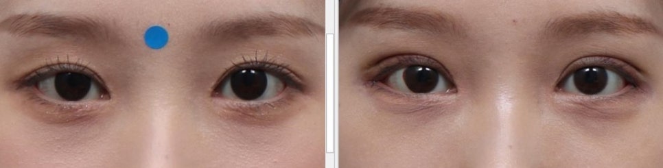 2. A dull eye shape due to ptosis
Ptosis refers to eyelid drooping caused by congenital or acquired factors. When the eyelid does not rise properly due to ptosis, part of the eyeball is covered, creating a dull eye appearance. In some cases, it refers to drooping of the upper or lower eyelid, but most commonly it is the upper eyelid that droops.
In this case, the most suitable surgical method is to increase the contraction strength by pulling or folding the levator aponeurosis and Müller’s muscle separately or together through an anterior approach (skin incision).
A simple self-test can be used to check for ptosis. After fixing the eyebrow with a finger, close your eyes and then open them. If the eyes open easily, it is negative; if they do not open easily without strength, it can be considered positive.
In this patient’s case, the problem was clearly identified in the photos. In the photo, the left side has a drooping upper eyelid so the eye light is not visible, while on the right side the eye light is visible. Therefore, through an anterior approach (skin incision), ptosis correction and incisional double-eyelid surgery with lateral canthoplasty were performed, successfully improving the ptosis to within 0.1 to 0.2 mm.
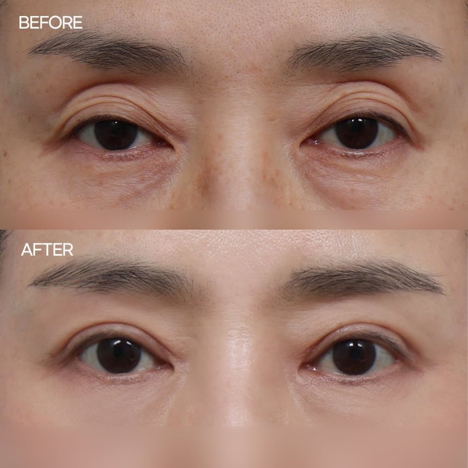 3. Sunken eyes and double eyelid folds caused by sagging skin (middle-aged plastic surgery)
In middle-aged plastic surgery, aging causes a loss of elasticity and the skin tissue sags downward. At the same time, the upper eyelid also droops, causing eye fatigue, and one may frequently strain the forehead or eyebrows to lift the eyelids, which can deepen wrinkles.
In this case, after performing an anterior approach (skin incision) with upper blepharoplasty, some unnecessary tissue is removed depending on the degree of sagging, and the sagging is improved with reinforcement using the orbicularis oculi muscle (the facial muscle surrounding the eyes). Sunken areas can be improved through fat repositioning above the eyelid.
If sunken areas still remain later, more delicate correction is possible by injecting filler into the preorbital space beneath the orbicularis oculi muscle through supraorbital filler.
In this patient’s case, upper blepharoplasty + upper eyelid fat repositioning + ptosis correction + reinforcement using the orbicularis oculi muscle improved the drooping eyelid and sunken areas, and at a later follow-up, since some sunken area remained, it was improved with 0.2 cc of supraorbital filler.
※ This case was used with the patient’s personal consent regarding portrait rights for a medical column.


#눈유형 #쌍꺼풀수술 #쌍수 #안검하수 #중년성형 #상안검수술 #바바성형외과