Every time you look at a photo taken from the side,
have you ever noticed that your lips stick out slightly,
and deleted the photo without even realizing it?
If you have been concerned about this for a long time, today’s post may be helpful.
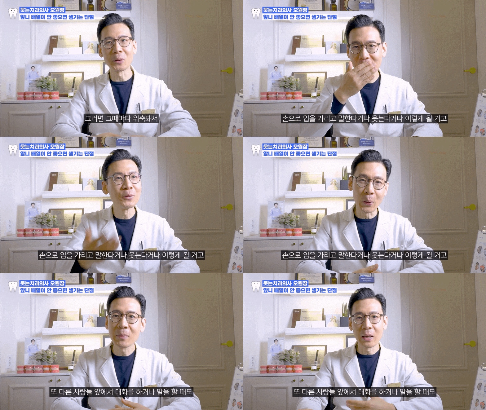

We would like to share the story of orthodontic treatment for gaps between the front teeth in a female patient in her 20s who visited Smile D Dental Clinic.
This patient was in her 20s,
and had been considering treatment for gaps between her front teeth for a long time because her front teeth were spaced apart and her lips appeared to project forward.
While searching Naver Blog for reviews of treatment for gaps between the front teeth,
she learned about Smile D Dental Clinic and began treatment after visiting the clinic for a consultation.
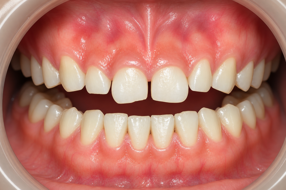
This image was created for illustrative purposes.
Detailed Diagnostic Process
After the visit, a panoramic X-ray and a lateral cephalometric X-ray were taken.
The patient’s concerns were gaps between the front teeth and protrusion,
and detailed analysis confirmed skeletal Class III malocclusion
along with facial asymmetry (HA).
This was not simply a dental issue.
Because skeletal factors and left-right asymmetry were also involved,
it was a case that required a careful approach from the diagnostic stage.
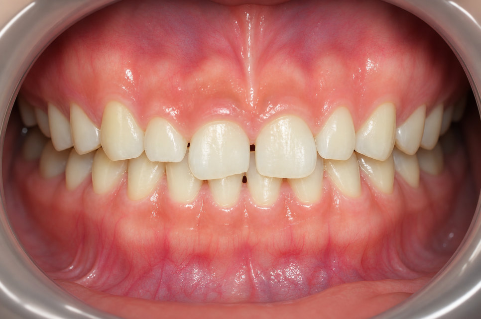
This image was created for illustrative purposes.
Why Treatment Was Performed Without Extractions
Even when asymmetry accompanies a skeletal Class III condition,
careful analysis of the available space in the dental arch and the range of possible tooth movement
can show that sufficient improvement is possible without extractions.
Detailed diagnosis indicated that this patient could improve the spacing and protrusion of the front teeth without extractions,
and that the molars could be finished in a Class I occlusal relationship.
Therefore, orthodontic treatment was performed without extractions.
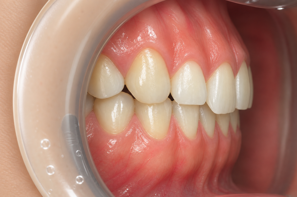
This image was created for illustrative purposes.
Whether extractions are needed is determined individually by considering the degree of protrusion, available space in the dental arch, and skeletal condition as a whole.
When, as in this case, good results can be expected without extractions,
it is my treatment philosophy to avoid unnecessary extractions.
< Smile D Dental Clinic Location >
< More About Smile D Dental Clinic >
| 📞 Smile D Dental Clinic |
|---|
| 0507-1449-2087 |
This image was created for illustrative purposes.
Treatment Plan for Gaps Between the Front Teeth and Orthodontic Appliances
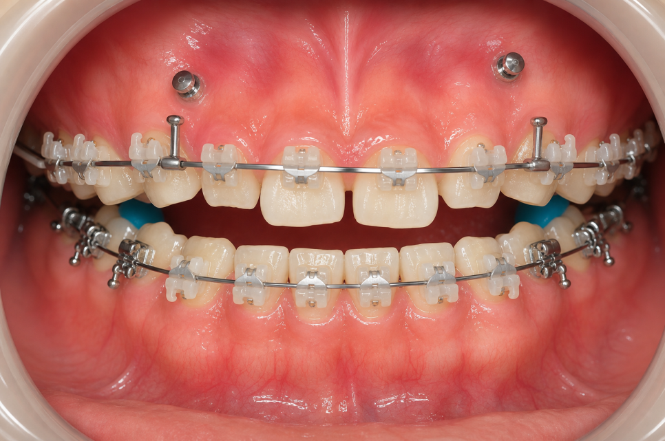
Treatment began with Clippy C (self-ligating appliances).
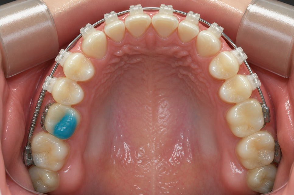
Two mini-screws (TADs) were placed in the upper jaw to precisely control the position and angle of the front teeth.
Mini-screws serve as anchorage, helping the desired teeth move accurately without the molars being pushed out of position.
This image was created for illustrative purposes.
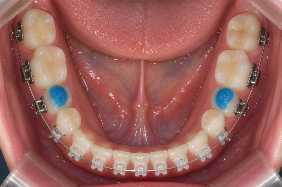
In addition, bite blocks were used for a certain period during treatment
to help the teeth move smoothly without occlusal interference.
This image was created for illustrative purposes.
In skeletal Class III cases,
an important treatment goal is not only to close the gaps between the front teeth,
but also to stably establish a Class I occlusal relationship in the posterior teeth.
Treatment Process for Gaps Between the Front Teeth
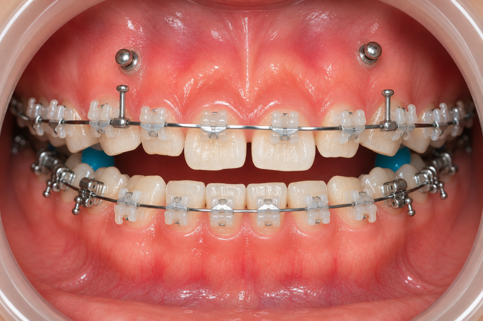
Treatment Process ① Appliance Placement and Initial Alignment
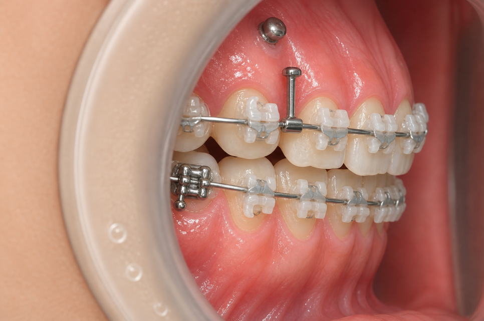
The initial stage focuses on evenly arranging the teeth.
The entire dental arch was aligned by starting with a thin wire and gradually replacing it with thicker wires.
There was mild pain and a feeling of having something foreign in the mouth at first, but the patient adapted within two weeks in most cases.
This image was created for illustrative purposes.
Treatment Process ② Space Closure and Occlusal Adjustment
While closing the space between the front teeth,
occlusal adjustment was performed using mini-screws and bite blocks, taking the skeletal Class III characteristics into account.
Only the necessary teeth were moved in a focused manner without moving the molars,
and changes in the side profile and asymmetry gradually began to appear at this stage.
This image was created for illustrative purposes.
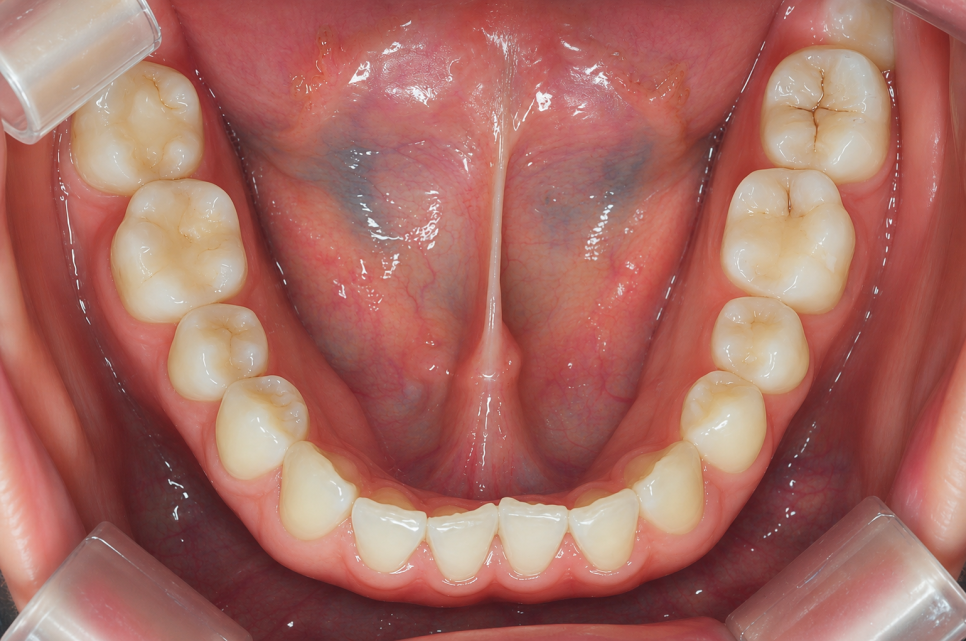
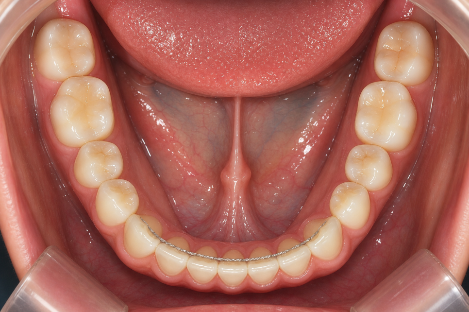
Treatment Process ③ Final Precision Adjustments and Appliance Removal
This was the final stage, involving fine adjustments to the tooth positions and occlusion.
After confirming that the posterior teeth were stably meeting in a Class I relationship, the appliances were removed,
and a retainer was fitted.
Previous image
Next image
This image was created for illustrative purposes.
Results After 1 Year and 2 Months of Treatment
After approximately 1 year and 2 months of orthodontic treatment,
the previously spaced front teeth were neatly aligned,
and the appearance of lip protrusion was naturally improved.
The posterior teeth were finished in a stable Class I occlusion,
and discomfort caused by asymmetry was also significantly reduced.
With the teeth properly aligned, brushing became much easier,
and the condition of the gums also visibly improved.

“It’s amazing that things could change this much without extractions.
It feels like 1 year and 2 months passed so quickly!”
If you have been putting off treatment for a long time because you are concerned about gaps between your front teeth or your side profile,
we recommend getting an accurate diagnosis at least once.
Whether extractions are needed, the treatment duration, and the type of appliance
vary depending on each person’s skeletal and dental condition,
so it is important to establish a treatment plan suited to you through an in-person consultation.

Located at Sinsa Station in Gangnam, Smile D Dental Clinic
establishes individualized treatment plans based on detailed cephalometric analysis
and follows the principle of treatment provided directly by an orthodontic specialist.
If you have similar concerns, please feel free to contact us for a consultation at any time :)
< Smile D Dental Clinic Location >
< More About Smile D Dental Clinic >
| 📞 Smile D Dental Clinic |
|---|
| 0507-1449-2087 |
※ This post is an informational post created by a medical institution in compliance with Articles 56 and 57 of the Medical Service Act and related medical regulations.
It was written solely on the basis of informational content exempt from review of medical advertisements.
The images are explanatory materials provided to aid understanding, and actual treatment is determined after consultation with medical professionals.