About Frontal bossing reduction
About Frontal bossing reduction
I am Dr. Kim Jun-hyun of Midline Plastic Surgery, where lifting is the focus and details make the difference.
People with prominent brow bones often worry that they look strong-faced,
and that the area above the brow bone (supraorbital ridge) appears sunken in profile.
If a forehead lift is performed and a flat forehead is improved,
this can partially address the problem.
However, for a more complete improvement, it is necessary to shave down the protruding bone.
With CT images and actual photos,
let me show you the patient I will introduce today.
The patient’s biggest concern was the strong-looking impression caused by the protruding brow bone,
and came to the clinic hoping to improve this.
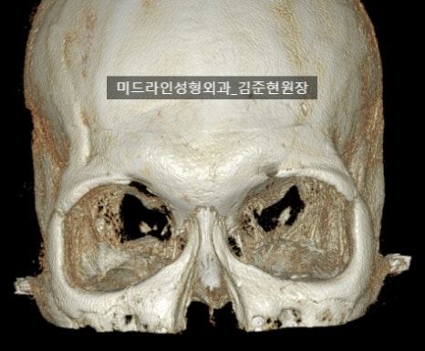
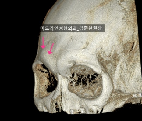
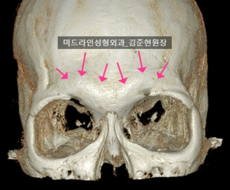
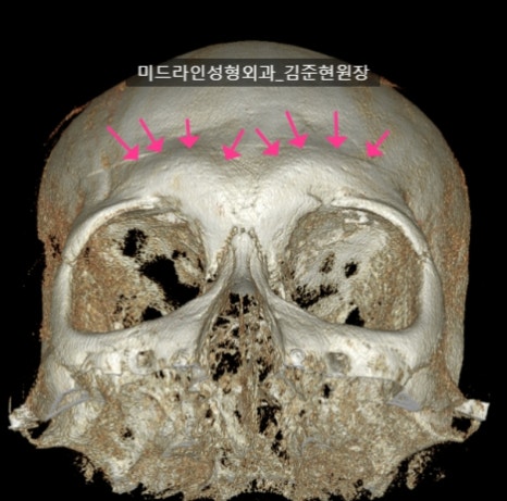
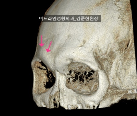
First, let’s look at the preoperative 3D CT.
You can see that the brow bone (frontal bossing) area is protruding.
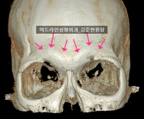
Preoperative CT: the prominent brow bone is observed at the purple arrow.
Before the shaving procedure, the CT shows the prominent brow bone at the purple arrow.


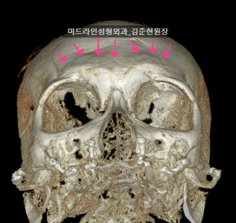
Preoperative CT: the prominent brow bone is observed at the purple arrow.
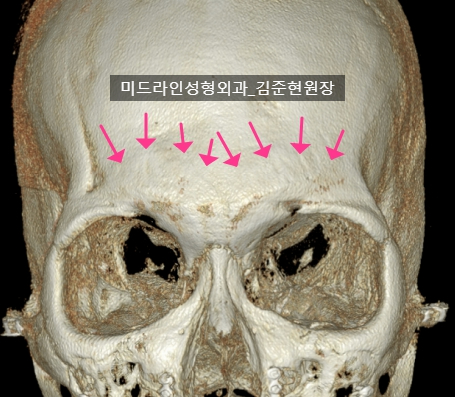
Depending on the angle, the contour of the brow bone can appear even more prominent.
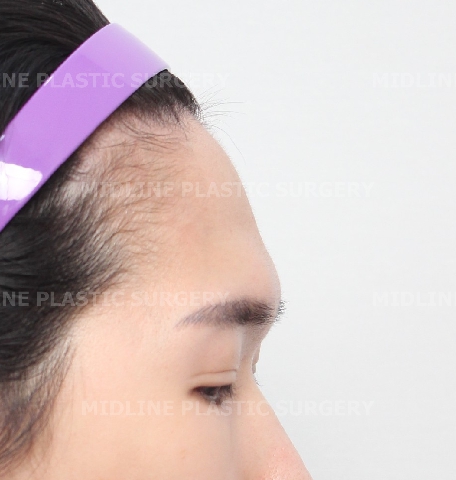
In real-life situations rather than on CT images,
especially when light falls on the face,
the brow bone contour can look more protruded,
which is why many patients say they want to improve it.

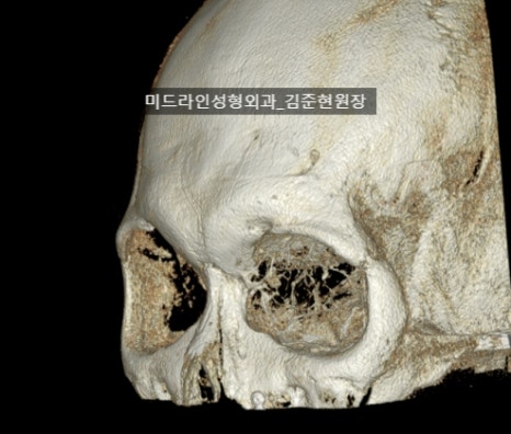
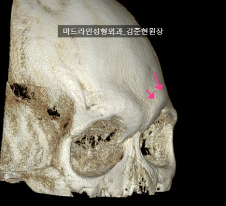
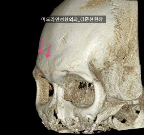
Preoperative CT: the prominent brow bone is observed at the purple arrow.
Even in the 45-degree preoperative CT image,
you can see the curvature of the forehead and brow bone.
Preoperative CT: the prominent brow bone is observed at the purple arrow.
As I mentioned in a previous post,
what must be carefully checked before shaving is the frontal sinus and the thickness of the bone covering it (the anterior table).
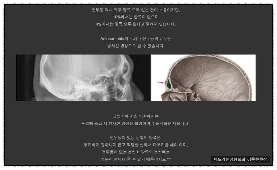
If you shave without checking the bone thickness,
the frontal sinus can be opened.
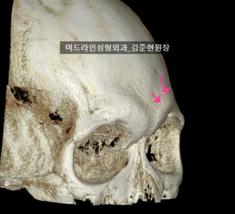
The patient who came this time had a large and wide frontal sinus, while the anterior table was thin.
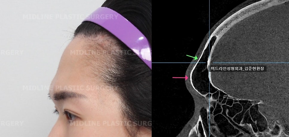
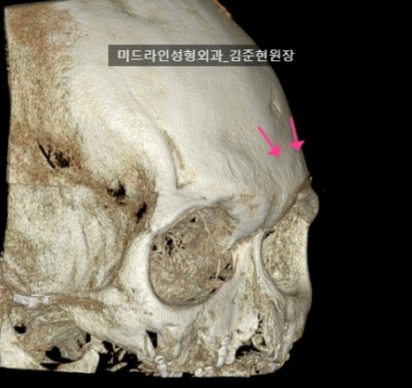
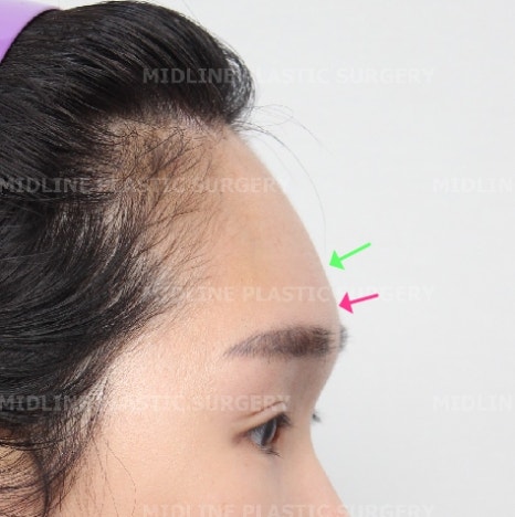
Side view and CT (sagittal view) of a person with a developed brow bone: you can see the thickness of the bone covering the frontal sinus.
This area must be checked when designing and before proceeding with surgery.
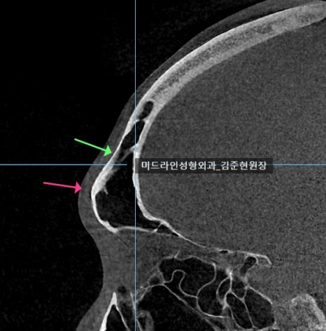
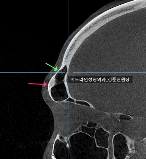
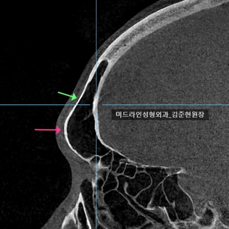
Depending on the location, some areas are thick and some are thin.
The area above the frontal bossing (fluorescent green arrow) is relatively sunken, and the bone there was the thinnest.
However, since this is not the area to be shaved, it is not something that requires much concern.
It is shaved while being viewed endoscopically.
The difference before and after surgery is checked with CT during follow-up.
Left) Before surgery / Right) After surgery
Left) Before surgery / Right) After surgery
Left) Before surgery / Right) After surgery
Left) Before surgery / Right) After surgery
The CT confirms that the protruding brow bone seen before surgery has become flatter.
Let’s also look at the postoperative photos.


As the prominent brow bone became lower,
the side profile became much softer.
This was a time to show that for people troubled by a protruding brow bone, brow bone reduction can be a good improvement option.
<If you have additional questions, please leave a private comment,
and I will kindly answer them. Thank you.>

At our clinic, when consulting for brow bone reduction,
we always take a CT scan.
The reason we must take a CT scan is to check
the thickness of the frontal sinus and the bone covering it (the anterior table).
If this is not checked, a representative complication of brow bone reduction,
namely opening of the frontal sinus,
can occur.
^^
.
.
.
※ If you add us as a neighbor, you can immediately read newly posted,
correct plastic surgery information. ※