The destiny of Secondary angle
in the advancement genioplasty
I’m someone who thinks advancement genioplasty is the highlight of contouring surgery.
That’s because the effect is so dramatic.
For people with a small chin:
- the jawline looks flat,
- a double chin can appear even if there isn’t much fat,
- the mouth looks relatively protruded.
These can become major sources of stress.
Advancement genioplasty is a procedure that can address all of these at once.
Even though advancement genioplasty can solve so many concerns, it is also true that patients naturally become more worried when preparing for surgery.
Among the questions I hear fairly often is one about the secondary angle.
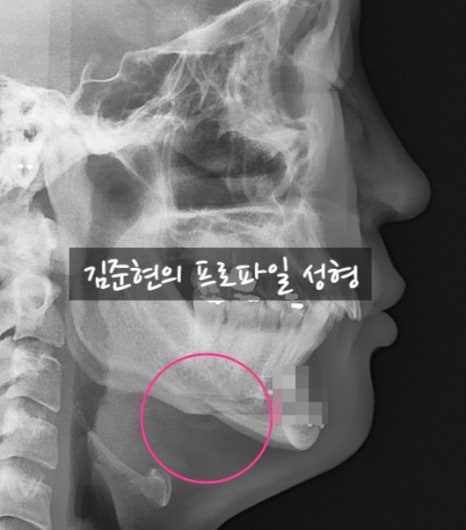
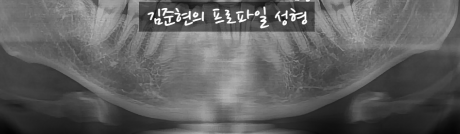
The circled part in the X-ray above
is the part referred to as the secondary angle or the submental step.
This was a patient who advanced 10 mm,
and this image was taken 2 years after surgery.
Let’s see how the secondary angle changed.
It can be seen that the line connects smoothly.
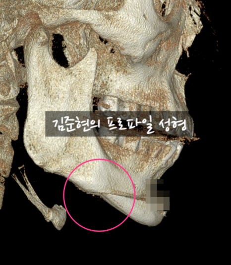
As bone union occurs, bone resorption takes place at the cut ends of the osteotomy.
Through this process, the result above is produced.
Therefore, if the secondary angle is shaved down during chin advancement,
the area will not connect naturally like in the X-ray and CT above,
and the shaved portion would end up appearing hollowed out.
On CT, it may seem like the bone should be shaved down and smoothed so the outer skin surface also looks smooth,
but in reality, that is not the case.
If anything, shaving the secondary angle can make it look depressed.
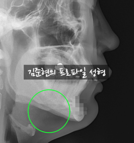
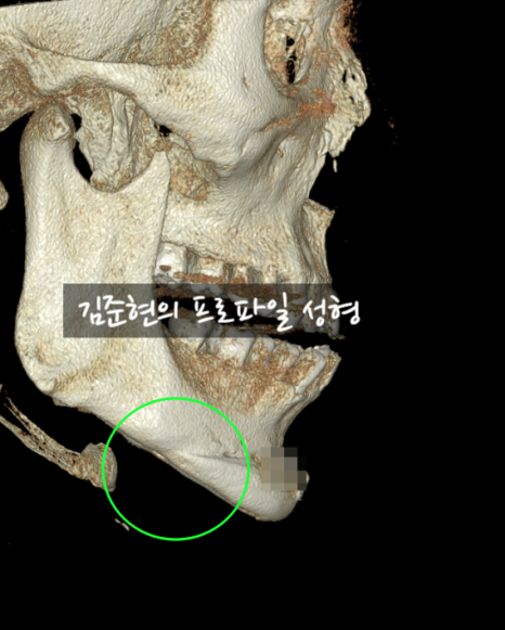
However, there is an exception here as well:
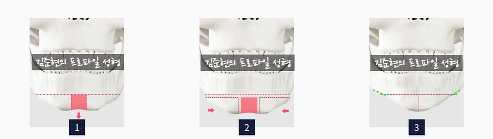
After T-shaped osteotomy, the secondary angle that forms is smoothed. Marked with a fluorescent light-green dashed line in Figure 3.
When performing a T-shaped osteotomy, if the width of the front chin becomes narrower, there is a difference in the width of the connecting bone,
and because this can be felt noticeably and may be visible on the surface,
only in cases of T-shaped osteotomy do we smooth both secondary angles.
I have prepared radiographic images before surgery, 7 days after surgery, and 2 years after surgery.
 Before surgery
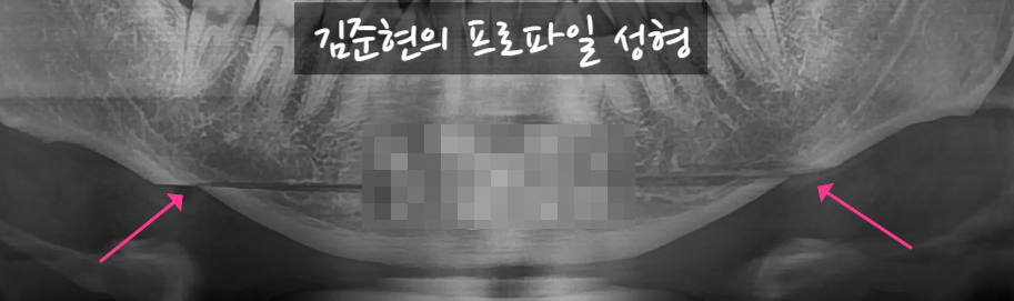 7 days after surgery, with the osteotomy site marked by a fluorescent magenta arrow.
 2 years after surgery, the previously cut area has become smooth. Marked with a fluorescent light-green arrow.
In the panoramic images above as well, it can be seen that the step at the osteotomy site
has become smooth over the course of 2 years.
In this way, the secondary angle seen on CT and X-ray after advancement genioplasty
improves to the point where it can be considered almost nonexistent.
So you can undergo advancement genioplasty with confidence.
<If you have additional questions, please leave a private comment,
and I will kindly answer them. Thank you.^->