Hello, I am Dr. Kim Jun-hyun, director of Midline Plastic Surgery Clinic. ^-^
Many people are interested in brow bone reduction.
So!
I would like to explain one of our clinic’s signature procedures, brow bone reduction:
how the consultation process works,
what we check before surgery,
how the most important aspect—the surgical result—turns out,
and what we compare when reviewing the progress.
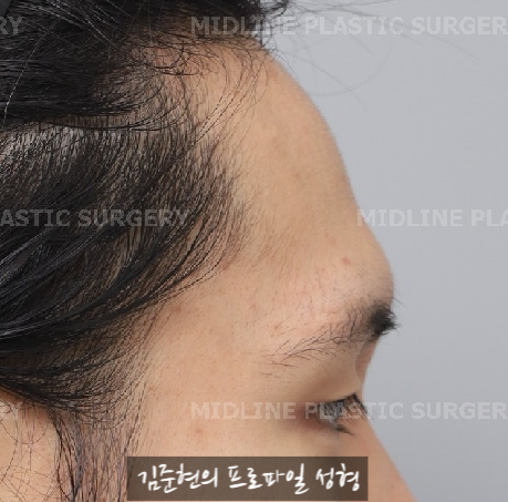

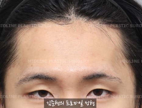
This person came in because they were concerned about their brow bones.
For a more detailed diagnosis, I recommend getting a CT scan starting at the consultation.
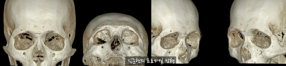
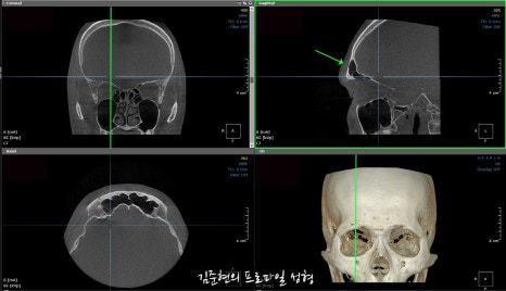
A 3D CT scan allows us to check the shape of the brow bones and, through the sagittal sections, determine the size of the frontal sinus and the thickness of the bone covering it at different locations.
That is why, if a CT scan was not taken during the consultation, it will be required when deciding on surgery.
This is the 3D CT scan taken during the consultation.
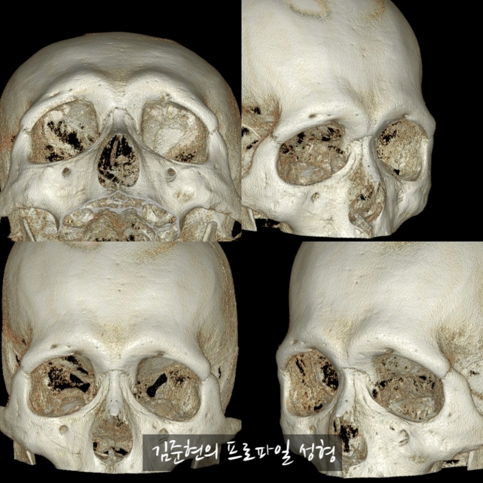
Before surgery, as shown here,
both the central and outer portions of the brow bones were significantly developed.
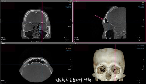
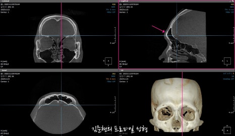
This is the preoperative sagittal-view CT scan.
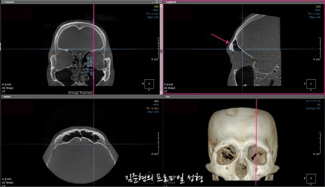
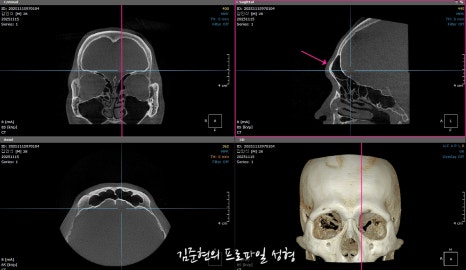
Left) Left side of the frontal sinus / Right) Center of the left side of the frontal sinus
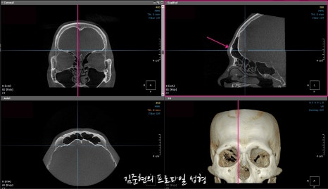
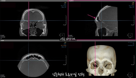
Left) Center of the right side of the frontal sinus / Right) Right side of the frontal sinus
The bone covering the frontal sinus is marked with purple arrows.
Because the bone was not thin, it could be sufficiently reduced.
In this way, the preoperative CT scan allows us to determine where and how much bone can be reduced.
This person did not want any change to the appearance of their eyes, so only brow bone reduction was performed.
Brow bone reduction can be performed on its own like this, and reducing the brow bones does not cause the eyelids to droop.
This is seven months after surgery.
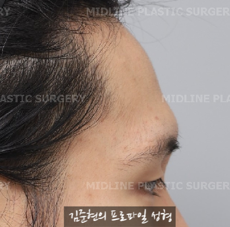

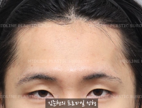

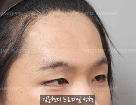
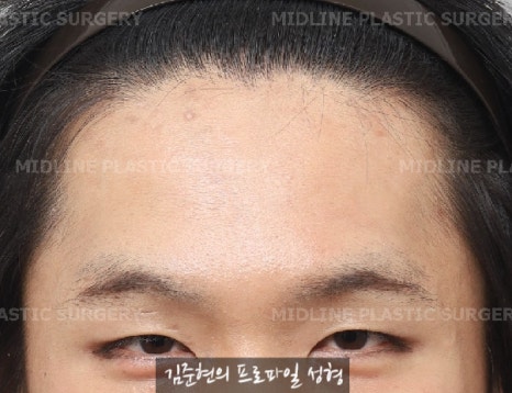
Top) Before surgery / Bottom) Seven months after surgery
As the brow bones that had been a concern were lowered, the person’s appearance became noticeably softer.
When reviewing progress, I capture and compare the 3D CT scans from before and after surgery.
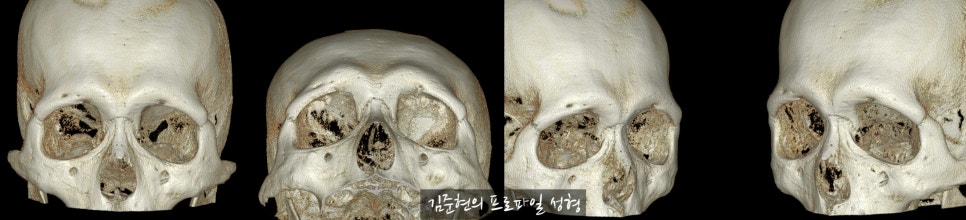
3D CT of the brow bones before surgery
3D CT of the brow bones after surgery
These are sagittal-section views.


They are small and difficult to see, so let’s enlarge and compare the left and right sides separately.
This is the area of the left frontal sinus.
Left) Left frontal sinus before surgery / Right) Center of the left frontal sinus before surgery
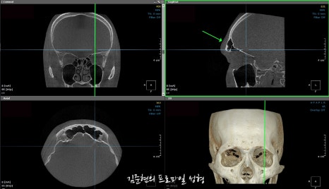
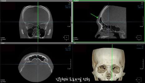
Left) Left frontal sinus after surgery / Right) Center of the left frontal sinus after surgery
This is the area of the right frontal sinus.
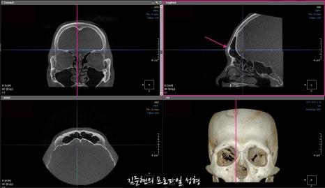
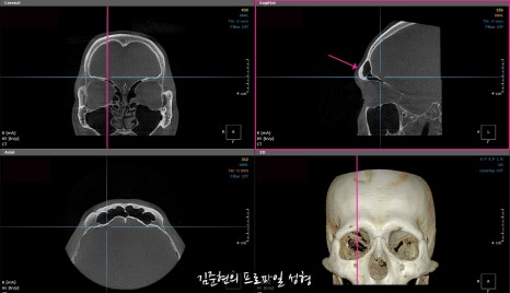
Left) Right frontal sinus before surgery / Right) Center of the right frontal sinus before surgery
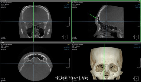
Left) Right frontal sinus after surgery / Right) Center of the right frontal sinus after surgery
When comparing the images above and below, you can see that the frontal sinus has not been opened, while the brow bones covering it have been effectively reduced.
This case was included because the person was very pleased that their brow bones had been lowered without changing the appearance of their eyes.
Brow bone reduction does not necessarily have to be performed together with a forehead lift.
It can also be performed on its own, so please feel free to contact us with any questions.
<If you have any additional questions, please leave them in a private comment, and we will be happy to answer them. Thank you. ^-^>