An osteoma is a benign tumor caused by the proliferation of osteocytes, and it is not a malignant tumor that affects health.
It commonly occurs on the forehead in the face, and when it becomes larger, it can look cosmetically unappealing, so many people look into having it removed.
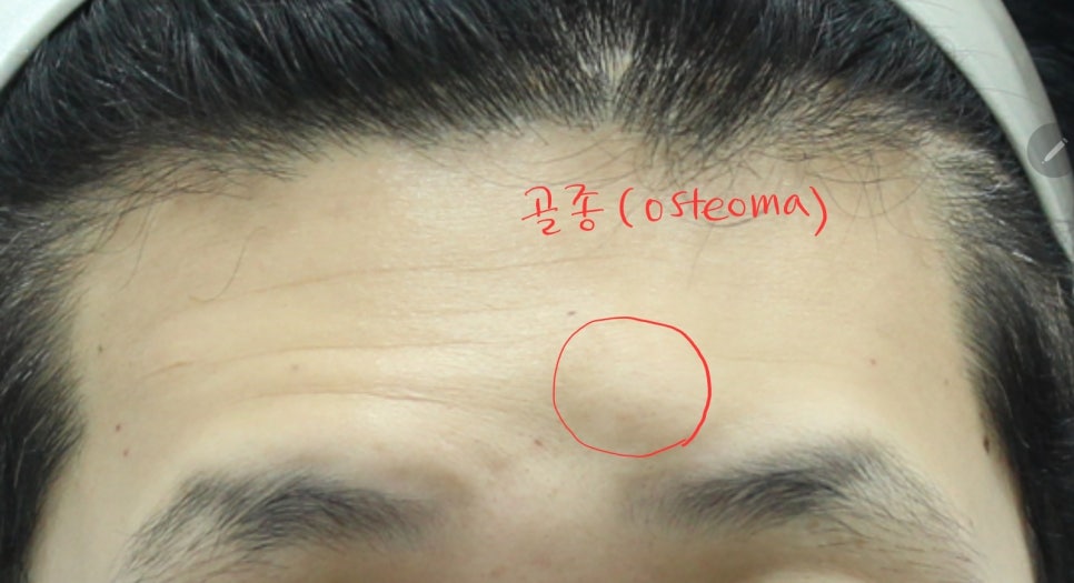
Osteoma on the forehead
In the preoperative examination, it is usually easy to diagnose an osteoma by touching the forehead and feeling the hard, bony texture, but a CT scan is performed for final confirmation.
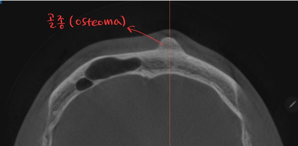
CT scan of an osteoma
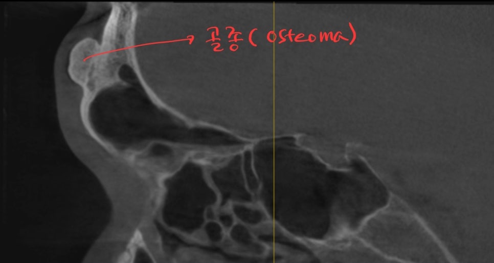
Side view CT scan of an osteoma
We confirmed an osteoma attached to the forehead bone, measuring about 1.5 cm to 2 cm in size.
In the past, osteomas were removed through a direct incision on the forehead, but this left an incision scar on the forehead, so recently the trend has been to avoid direct incisions whenever possible.
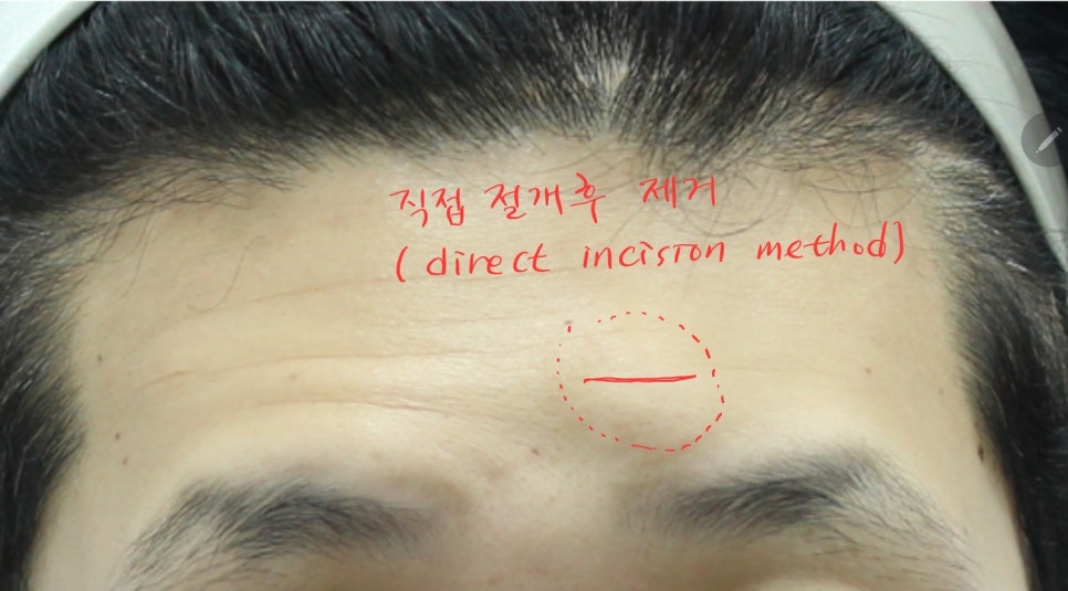
Past method of direct incision removal of osteomas
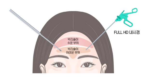
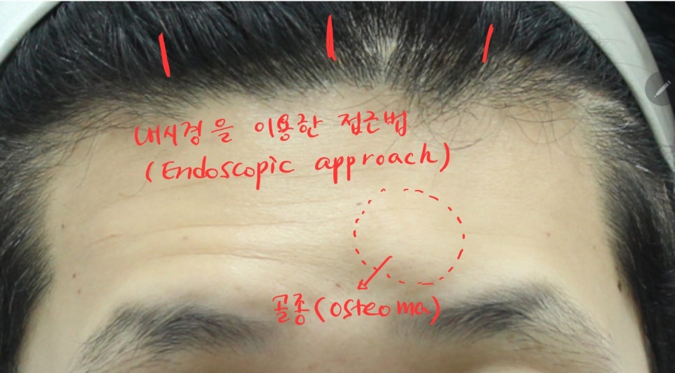
Recently, with the development of endoscopic surgical techniques, osteomas are being removed by approaching from inside the scalp.
In particular, if forehead wrinkles are present or eyelid drooping is accompanied, performing the procedure together with an endoscopic forehead lift can result in very high cosmetic satisfaction.
Dissection in an endoscopic forehead lift
Endoscopic forehead lift and osteoma removal
Using the same incisions as an endoscopic forehead lift, the forehead is dissected through small incisions in the scalp.
Because dissection is performed accurately through a high-definition endoscope, it can be done without damaging the nerves and blood vessels of the forehead.
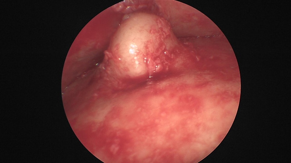
Forehead osteoma seen through the endoscope
The forehead osteoma is identified through the endoscope and removed using various surgical instruments, and the surrounding forehead bone is also carefully refined.
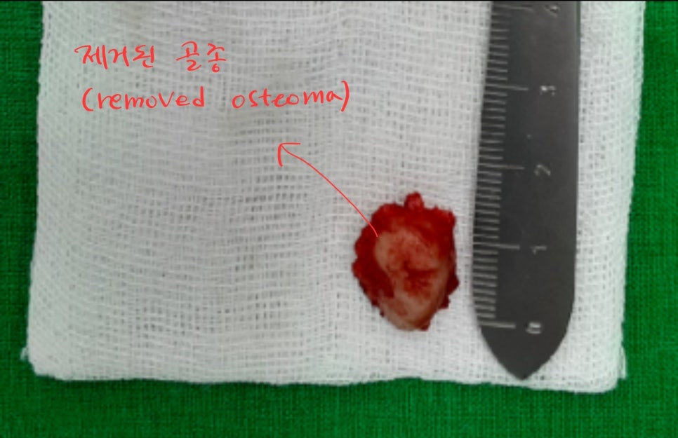
Forehead osteoma removed through the endoscope
After the osteoma is removed, the forehead lift procedure is performed in the same way.
The forehead and brow are lifted through separation of the periosteum, release of retaining ligaments, and radiofrequency treatment of the corrugator muscles.
Let’s compare before and after surgery.
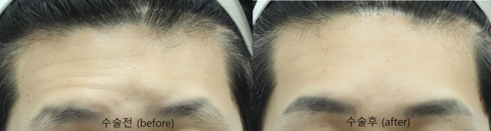
Front view comparison before and after surgery
The osteoma on the forehead has been removed, leaving the forehead smooth.
Through the forehead lift, forehead wrinkles have also been greatly reduced, and the asymmetry of the eyebrows and the sagging skin around the eyes have also improved.
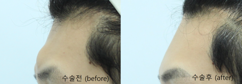
Side view comparison before and after surgery
From the side as well, the forehead osteoma has been removed, resulting in a smooth forehead shape.
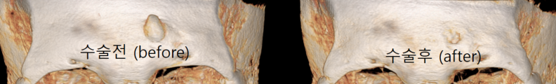
CT comparison before and after surgery
The CT images also show that the osteoma has been removed well. After the osteoma is removed, the removed area heals as callus forms. On CT, the callus appears with slightly lower density, and later, when enough bone cells fill in, the density on CT also becomes smoother.
In addition to osteomas, various benign tumors such as lipomas and epidermoid cysts can also occur on the forehead.
In the case of benign tumors that occur under the skin, there are many cases in which they can be removed together with an endoscopic forehead lift.
Surgery using an endoscope avoids a direct incision, so it does not leave a scar on the forehead, and it can also address forehead wrinkles and sagging skin around the eyes, so it may be worth considering.