
An impression that conveys trust and warmth
is often associated with clear, bright eyes
and a bright facial expression.
For this reason, many people undergo fat grafting
to eliminate the shadows in the tear troughs,
which are a major cause of a dull and tired-looking
eye area, but eventually
visit a clinic for
foreign-material removal under the eyes.
They expected to achieve
a brighter, more vibrant appearance,
but when the results were not as satisfactory,
they eventually sought medical attention
after much concern.
Whether it is filler or fat grafting,
once the material has taken hold,
it can be difficult to remove. Therefore,
it must be removed safely by
an experienced physician.

First, looking at the causes of tear troughs,
some people have prominent tear troughs
because they are born with little fat
and have weak surrounding tissue,
while others develop them due to
lifestyle habits.
If the procedure is performed without
understanding the cause, the tissue
at the grafted area may become uneven
and adhere abnormally, making
additional foreign-material removal
under the eyes necessary.
In younger people in particular, causes include
lack of sleep and stress, as well as
excessive computer and smartphone use.
The fascia around the eyes repeatedly contracts
and relaxes, losing its elasticity,
which causes the trough area to become hollow.
Blood circulation decreases, the blood vessels
become more visible, and the skin darkens,
making the issue appear even more pronounced.

Many people assume that this kind of aging change
only appears when they reach middle age,
like an elderly statesman,
so younger people in their 20s and 30s
often report considerable stress.
After undergoing fat grafting or filler
after much deliberation, they visit our clinic
for a consultation about
foreign-material removal under the eyes.
In fact, tear troughs and protruding fat
tend to become more severe with age,
but they can also appear sufficiently
in younger people, so the cause must be
examined first.
Based on anatomical knowledge,
we carefully assess the skin, septum,
ligaments, and orbital fat by tissue layer
and make a comprehensive diagnosis.
Fat grafting is performed by grafting
a person's own fat into hollow areas
under the eyes to create natural volume
and help brighten the appearance of
dark circles and a dark, tired-looking face.
In simple terms, balance is restored
through removal or grafting, depending on
whether the area is protruding or hollow.
Because the person's own tissue is used,
the risk of side effects is relatively low,
and the take rate is generally high.
However, if the fat is not evenly repositioned
or adheres unevenly after clumping to one side,
the area may become uneven.
Excessive grafting can also cause bulging,
and because selective correction can be difficult,
people seek foreign-material removal
under the eyes.

Immediately afterward, however,
this may occur naturally as swelling subsides
and the tissue settles into place.
If the condition remains unchanged
for two to three weeks,
or persists even three months later,
when the area should have fully stabilized,
it may be a significant issue requiring
appropriate treatment.
The same applies to filler:
lumps can form when too much is injected
or when it is not distributed evenly.
In some cases, it may also move with
muscle movement and create unnecessary lumps,
or absorb moisture within the tissue
and swell.
The skin around the eyes is very thin
and contains many blood vessels,
so filler is often not distributed evenly.
It must therefore be injected delicately
using an appropriate technique.
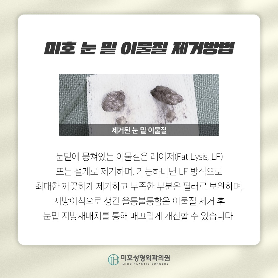
When the area is examined with ultrasound
before foreign-material removal under the eyes,
cases in which the filler was placed
in an incorrect location are also found.
Absorbable filler is naturally absorbed
over time, whereas permanent filler
does not disappear and must be removed physically.
Even semi-permanent filler can irritate
surrounding tissue and create scar tissue
before it disappears. After six months,
any remaining material may show the characteristic
of being absorbed and leaving residual tissue.
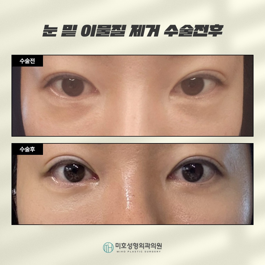
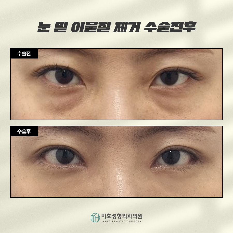
Therefore, when the results of fat grafting
or filler are unsatisfactory—for example,
when the material appears uneven,
has taken hold in granules, or has swollen—
it is possible to improve the appearance
toward the originally desired result
by removing it thoroughly and performing
under-eye fat repositioning at the same time.
When considering foreign-material removal
under the eyes, caution is needed regarding
advertisements claiming that only the grafted fat
can be dissolved while the original fat tissue
is preserved, or that the material can be
removed indiscriminately by suction.
The location must first be identified
with ultrasound, and the material must be removed
carefully so that none remains in the body.
At our clinic, we begin with the understanding
that each person's physical condition
and characteristics are different.
By analyzing the tissue layer by layer,
we tailor and perform the anatomically necessary
procedures in detail.
Of course, the information shared
during the consultation is also actively reflected
in the treatment plan.
Depending on various physical characteristics,
we may use nano-fat grafting and fat repositioning
when the skin layer is thin; fat removal and
repositioning for contours protruding due to
orbital fat; or reinforcement with nano-fat grafting
and filler. Each person and each case
is approached differently.

Foreign-material removal under the eyes
is performed under local or twilight anesthesia,
though the method may vary slightly
from person to person.
An incision is made in the inner conjunctiva
to first remove the foreign material
and ligament adhesions.
After performing fat repositioning,
the septum is reinforced and the fat is secured
to complete the procedure in a stable condition.

It is gratifying to see the concerns of people
who were often told that they looked tired
because the area under their eyes was bulging
and dark become resolved through fat repositioning.
Because it can provide natural improvement
and a high level of satisfaction with life,
it is a method that many people consider
at least once.
Since the skin in this area is particularly thin
and foreign-material removal must be performed
close to the skin under the eyes,
it is important to proceed carefully and safely.
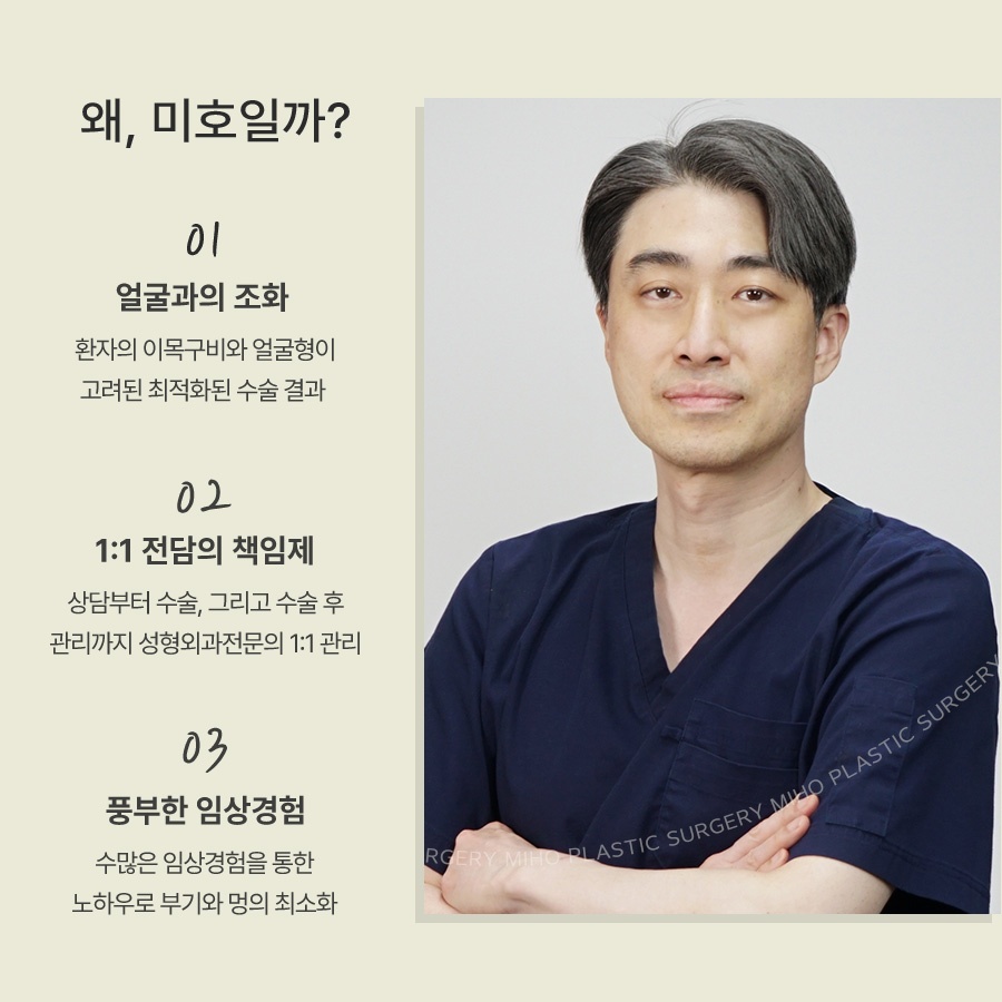
Be sure to choose a trustworthy institution
with extensive clinical experience,
thorough diagnosis and follow-up care,
and a physician-name disclosure system.

Previous image
Next image