
As interest in improving the shape of the nose grows,
more people are also considering which graft material to use.
In particular, an increasing number of people feel hesitant about
artificial implants such as silicone or Gore-Tex,
or are considering new materials when undergoing another procedure
after having already undergone nasal contour improvement once.
Against this backdrop, autologous costal cartilage rhinoplasty
is frequently mentioned as one option.
Today, we will take a step-by-step look at
how this approach, which uses the patient’s own tissue,
is performed and what the recovery process is like.
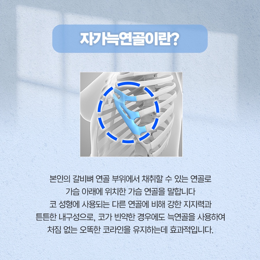
Autologous costal cartilage rhinoplasty involves
harvesting part of the cartilage at the end of a rib
and using it as material to shape the structure of the nasal contour.
Costal cartilage supports the flexibility of the rib cage
and helps cushion movement.
Because the material is obtained from the patient’s own body,
it is often chosen because the likelihood of a foreign-body reaction
or rejection is considered low.
In addition, unlike artificial materials,
the risk of the skin becoming thin or the material protruding through it over time
is significantly lower, which is another characteristic feature.
Its low absorption rate is also considered an advantage,
as it helps the shape remain intact for a long time.

There are various situations in which autologous costal cartilage rhinoplasty may be considered.
Typical examples include cases in which contracture, a representative complication,
occurs after a previous nasal contour procedure,
causing the nose to become shorter or appear turned upward,
as well as cases involving inflammation or a foreign-body reaction
caused by an artificial implant.
Costal cartilage may also be used as an alternative
when the nasal bridge is severely low from birth,
or when the support provided by cartilage harvested from the ear
or nasal septum is considered insufficient.
It can also be useful when part of the septal tissue was damaged
during a previous contour-improvement procedure
and the internal framework needs to be rebuilt.
In other words, it is often selected
when it is difficult to expect a sufficient result
using other materials.
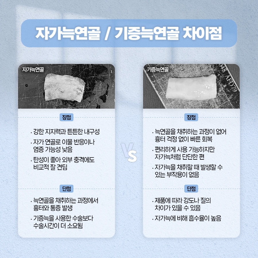
Compared with other autologous tissues,
the characteristics of costal cartilage become even more apparent.
Ear cartilage is soft and elastic,
so it is used to create a natural shape at the tip of the nose,
but it has limitations when strong support is needed.
Septal tissue, on the other hand, is flat and provides good support,
but the amount that can be obtained is limited.
Costal cartilage is harder and can be obtained in greater lengths
than other autologous materials,
so it can be used broadly in areas that require a supporting structure.
Its relatively strong resistance to inflammation
is also considered one of its main advantages.
Thanks to these advantages, this tissue can be used in various areas of the nose.
Most commonly, it is used for the strut that raises and supports the nasal tip
and for the columella, the area between the nostrils.
Depending on the needs of the individual,
it can also be used to reinforce a low nasal bridge.
It may be thinly sliced and refined
to serve as a surface material that naturally smooths out irregularities.
Another widely known method involves finely mincing it
and shaping it together with fascia or a biological adhesive
to create the nasal bridge line.
Because it can be processed in various forms in this way,
it can be designed flexibly according to the individual’s condition and circumstances.
Autologous costal cartilage rhinoplasty generally proceeds in three stages.
First, the required amount of cartilage is harvested
from the lower part of the rib cage.
The cartilage is then shaped into pieces or molded
according to its intended purpose.
To reduce warping, in which the material bends over time,
the central portion of the cartilage may be used preferentially,
or techniques may be used to shape the pieces symmetrically.
Finally, the prepared pieces are grafted
and shaped to create the desired contour.
Because the procedure is generally performed under general anesthesia
or deep sedation,
an anesthesiology team also participates.
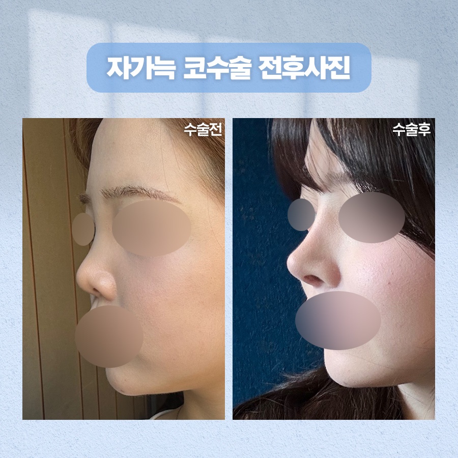
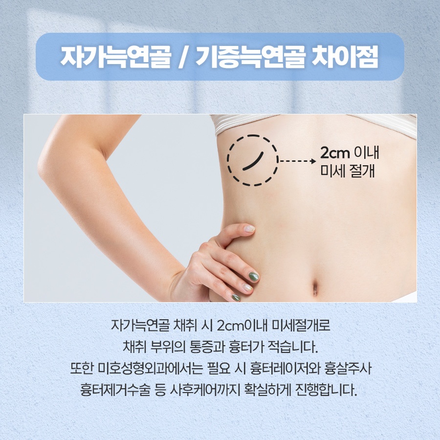
The cartilage is usually harvested
from around the sixth to ninth ribs,
and the incision is generally about 2–3 cm long.
To make the scar less noticeable,
the incision is often placed along the crease beneath the breast.
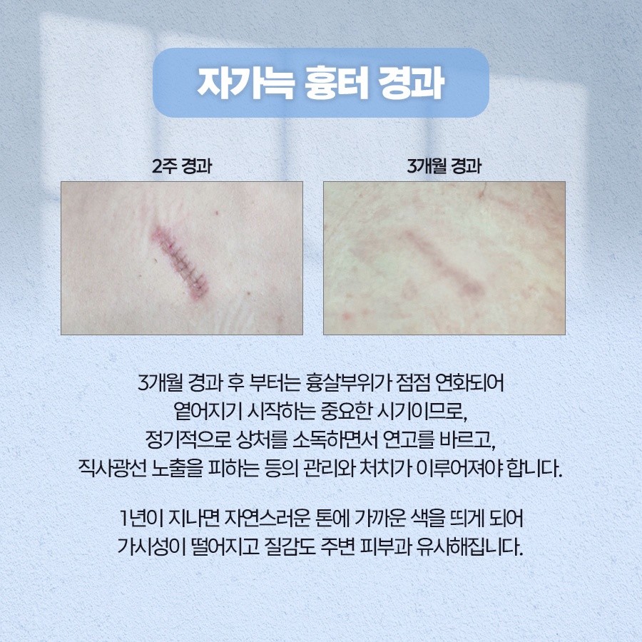
A waterproof bandage is applied to the harvest site,
followed by ongoing care using scar-reducing ointment.
The wound must also be closed carefully in multiple layers
for smooth healing.
The incision gradually becomes lighter over time,
but the extent to which a mark remains may vary
depending on individual skin characteristics.
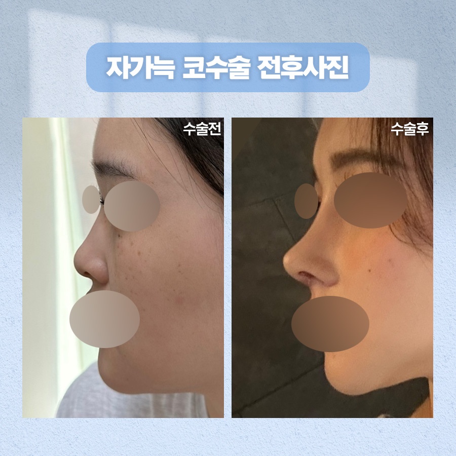
Next, let us look at recovery and care
after autologous costal cartilage rhinoplasty.
During the first 2–3 days, swelling and bruising may increase,
then gradually subside over time.
Cold compresses can be helpful during this period.
When sleeping, keeping the upper body elevated above heart level
can help manage swelling.
After approximately one week,
bruising and swelling generally subside considerably,
making it possible to return to everyday activities.
A compression band may be worn
to reduce pain at the rib harvest site
and help prevent an internal hematoma.
It is safer to avoid strenuous exercise
for about a month.
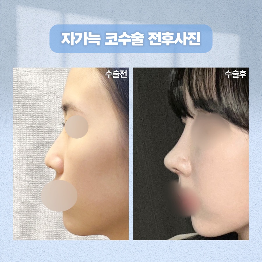
There are also points that require attention.
As mentioned earlier, warping
refers to an issue in which the grafted area
gradually bends over time,
which may lead to subtle changes in the nasal bridge.
In addition, because of the characteristics of autologous material,
some of it may be absorbed,
and chronic discomfort near the ribs
has also been reported in rare cases.
Furthermore, cartilage may undergo calcification and harden like bone with age,
so thorough evaluation beforehand is necessary.
Problems such as pneumothorax, in which air accumulates inside the chest,
may also occur during the harvesting process.
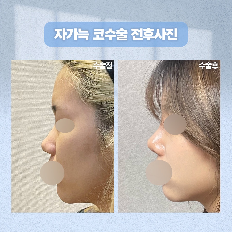
In summary, autologous costal cartilage rhinoplasty
may be a meaningful option when strong support is needed
or when using artificial materials is difficult.
However, it is not equally suitable for everyone,
and the outcome may vary depending on factors such as
an individual’s anatomy, skin thickness, and medical history.
The process of determining the design direction,
while also considering the desired appearance
and harmony with the proportions of the entire face,
has a significant effect on the quality of the final result.
Therefore, it is important to carefully assess your condition
through a consultation with a medical professional,
discuss the choice of material sufficiently,
and then make a cautious decision.

Previous image
Next image