After nose surgery, if some time passes and you happen to get an X-ray or CT scan, it is common to feel worried when you see an unexpected result. In particular, when the nose silicone does not look straight and instead appears bent or seems to have shifted position, it is natural to become anxious about whether revision surgery is needed.
However, what appears on an image does not always match the actual condition of the nose. In this article, we will calmly look at why silicone can appear different on an X-ray taken after nose surgery and in what cases there is no need to worry.
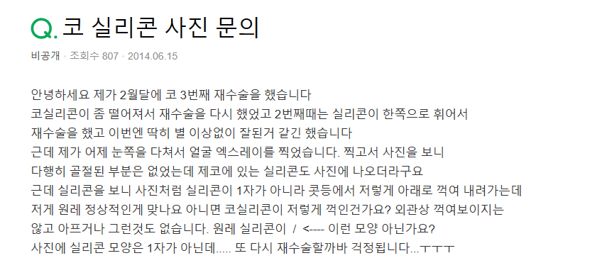
Q.
Hello. I had my second revision surgery this February, and I have had nose surgery a total of three times so far. The first revision was done because the nose silicone had moved down slightly, and the second revision was performed because the nose silicone appeared to be bent to one side. After surgery, I was doing well without any major issues, but recently I injured the area around my eye and had a full facial X-ray taken.
There was no fracture, but when I looked at the image, the nose silicone stood out, and instead of looking straight, it seemed bent around the bridge of the nose, which worried me. I am wondering whether this shape is a normal position or whether the silicone has become deformed. I do not have pain or any visible shape abnormality, but after seeing the image, I am anxious that I may need revision surgery again.
A.
When a CT or X-ray is taken after nose surgery, it is relatively common for the nose surgery silicone to appear slightly bent on the image. This can happen because of the weight of the soft tissue over the silicone and the structural characteristics of the nose. The upper part of the silicone sits on bone, and as it extends downward it rests on cartilage, so on an X-ray it may appear bent, just as you saw.
If there is currently no pain and no visible change in shape, the possibility that this is a form abnormality or a problem is low, and in such cases you do not need to worry about revision surgery. If there are no issues with appearance or symptoms, you do not need to pay too much attention to it.
#NoseSurgerySilicone #KowonPlasticSurgery
This image was used with the model’s consent.
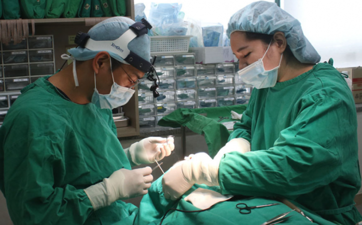
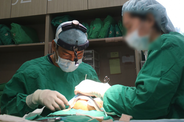
I am Kim Hyung-taek, the director of Kowon Plastic Surgery in Sinsa, focusing on a wide range of rhinoplasty procedures, including nose surgery using autologous rib cartilage. Today, I will introduce the revision nose surgery process for a female patient with multiple previous surgeries, focusing on a relatively difficult case that has received many inquiries in the Gangnam area.
This image was used with the model’s consent.
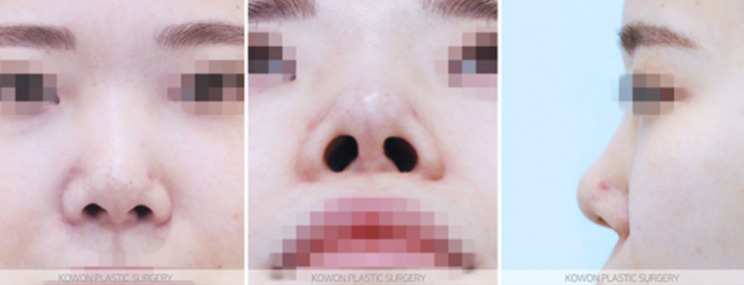
This patient had her first nose surgery about 7 years ago using donated rib cartilage, ear cartilage, and silicone, and later underwent one additional reconstructive surgery, but she returned because she was not satisfied with the results.
The hump on the bridge had not been sufficiently corrected in the previous surgery, so the bridge line was not smooth, and the nose also appeared shorter overall, which was another concern. For these reasons, this case could serve as a reference for those considering revision nose surgery.
This image was used with the model’s consent.
After examining the condition through Kowon’s consultation, the bridge still had irregularities in the middle, making the line look unnatural, and slight asymmetry in the nostrils was also observed.
Since she already had two previous surgeries and donated rib cartilage had been used in the first surgery, we recommended using autologous rib cartilage for a more stable result. This method is considered a relatively stable option in difficult female rhinoplasty cases.
This image was used with the model’s consent.
The patient wanted a slightly more striking impression while still keeping a natural look, so we planned to smooth the bridge and hump area with osteotomy so that the overall line would appear sharper. For the bridge, instead of the existing implant, we used rib cartilage particles in a way that considered both stability and naturalness.
This image was used with the model’s consent.
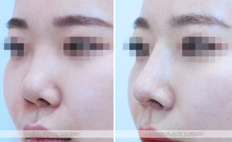
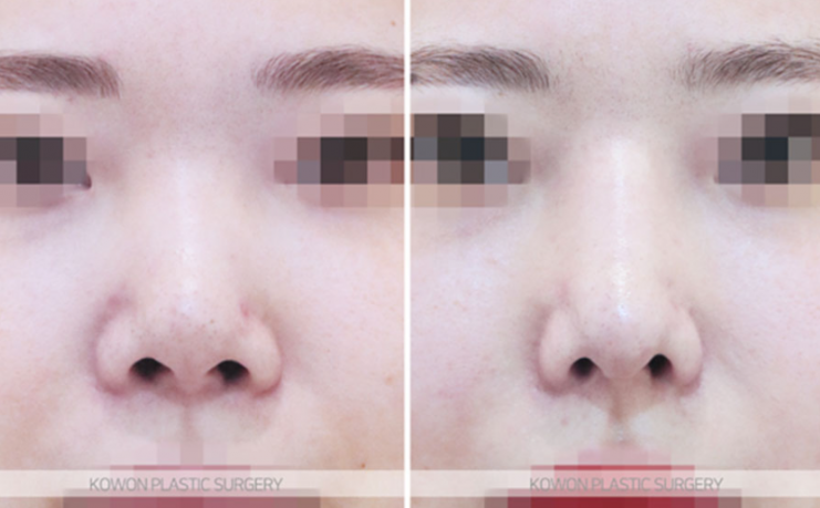
Looking from the front after surgery, the width of the bridge was clearly reduced, and the nose tip seen from below was also refined in height and shape according to the patient’s wishes. In particular, the nose tip line became cleaner, giving the overall impression a more sophisticated appearance.
This image was used with the model’s consent.
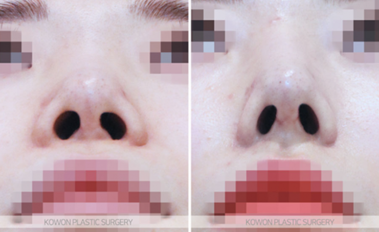
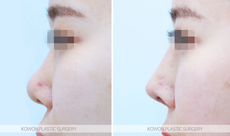
After surgery using autologous rib cartilage, the line from the glabella to the nose tip became straighter and smoother, and the middle part of the bridge that had previously looked hollow was also naturally improved.
From the side, the improvement in the nasolabial angle was clearly visible, which is one of the important elements in female rhinoplasty. At first, the bridge may appear somewhat high, but this was the result of designing with the expectation that some resorption would occur over time due to the characteristics of autologous tissue.
This image was used with the model’s consent.
This photo was taken about one month after surgery, and as the resorption and tissue stabilization process continues over the next six months or so, the line will settle into an even more natural shape.
As in the case introduced today, even when concerns have deepened after multiple surgeries, stable results can still be expected through sufficient consultation and planning. If you are considering revision nose surgery or improving the line of your nose, please visit for a consultation to receive detailed guidance.
A post worth reading for reference if you are worried about revision nose surgery
is "Q. In Gangnam, is this a nose tip surgery complication that requires revision nose surgery?"