Hello!
I am the director of Seoul Bardi Dental Clinic.
▼ Check the dental clinic location ▼
| This post was written directly by Seoul Bardi Dental Clinic for the purpose of providing medical information, in compliance with Article 56, Paragraph 1 of the Medical Service Act. The information provided is for reference only, and we recommend visiting a medical institution and receiving guidance from a medical professional for an assessment of your symptoms and accurate care methods. All procedures and surgeries performed at a dental clinic may involve risks depending on the individual, such as inflammation, bleeding, and swelling. Please make your decision after having a sufficient consultation with a medical professional. The treatment cases in this post were photographed with the consent of patients who completed treatment at our clinic, and the before-and-after photos were taken under identical conditions. |
| Directions to the Clinic |
Many people consider taking dental impressions
to be one of the most unpleasant procedures
at a dental clinic.
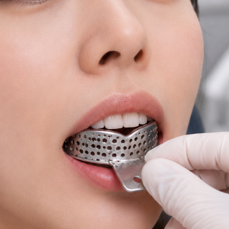
Example photos
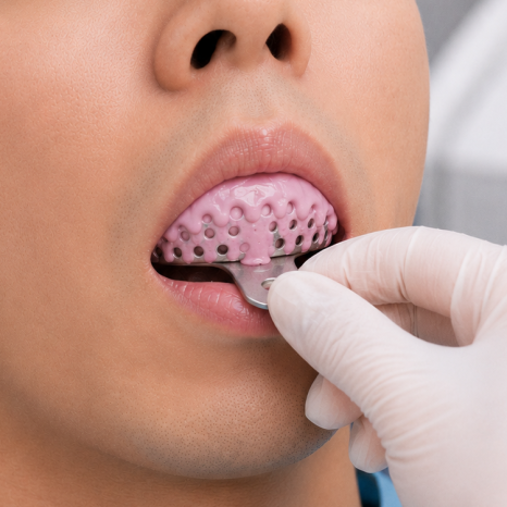
You have to bite down on material that hardens
while filling your mouth and endure it
for several minutes,
so you may have experienced gagging
or wondered, “When will this be over?”
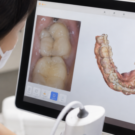
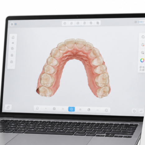
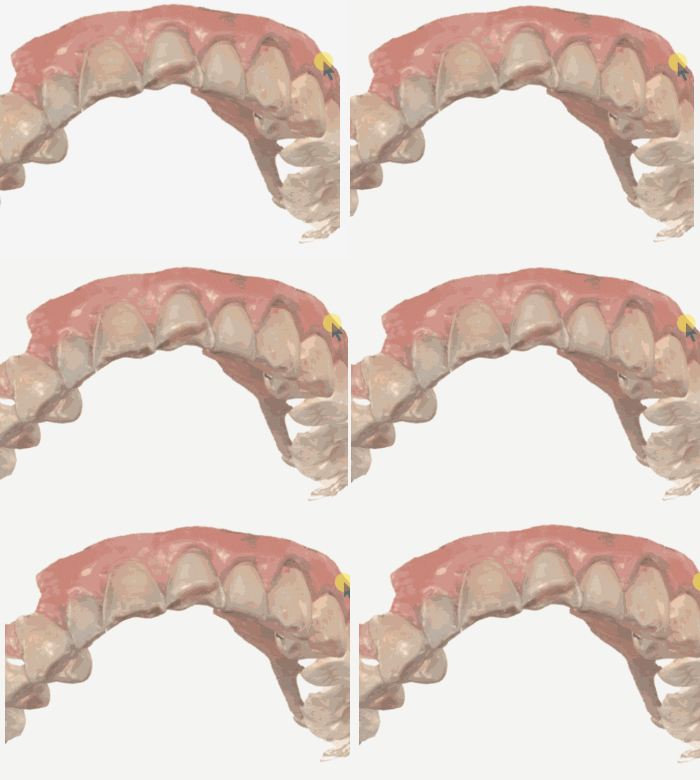
These days, an intraoral scanner can replace this process
by scanning the mouth like taking pictures with a camera.
Beyond simply making the process more comfortable,
it can also make a difference in accuracy.
Today, I will explain how digital impressions differ from traditional impression-taking,
where they offer advantages, and what limitations they have.
| Chapter 1. Why Did Errors Occur with Traditional Impression-Taking? |
Example photos
Traditional impression-taking involves
placing impression material in a tray,
having the patient bite down on it,
and waiting for it to harden.
There is a high possibility of errors occurring during this process itself.
Even a slight difference in the patient’s biting force or position
can distort the shape,
and areas obscured by saliva or bleeding
may not be captured accurately.
In addition, the material may shrink or deform
while it hardens and is transferred
into a physical model.
One of the advantages often cited for digital impressions
is precisely that they have a lower risk of deformation
during the impression-taking and model-production stages.
| Chapter 2. Are Intraoral Scanners More Accurate? — It Depends on the Case |
This is probably what you are most curious about.
To answer honestly,
digital impressions are superior for single-tooth restorations.
When producing a single crown,
research has found that digital impressions provide
significantly better marginal fit (the boundary between the tooth and restoration)
and internal fit than conventional silicone impressions.
(Systematic review, 2024)
This means that the restoration
fits the tooth more precisely,
which is advantageous for reducing the risk of
secondary cavities developing through gaps
or the restoration becoming dislodged.
However, the limitations are also clear.
When scanning a wide area at once,
such as a partially edentulous area where several teeth are missing
or a completely edentulous mouth where all teeth are missing,
accuracy may decrease.
(Umbrella review, 2024)
Some reports indicate that conventional impressions
may be more accurate in these cases,
so it is appropriate to use or select the two methods together
depending on the situation.
In other words, it is not accurate to say that
“scanners are always more accurate.”
They have advantages for restorations involving one or a small number of teeth,
while caution is needed when scanning a broad edentulous area.
| Chapter 3. The Biggest Difference Patients Notice: Comfort and Time |
Apart from accuracy,
the change patients notice immediately is comfort.
Taken together, multiple studies consistently show that
intraoral scanners reduce procedure time
and improve patient comfort
compared with conventional impressions.
(Umbrella review, 2024)
Since there is no need to bite down on material
and endure it for several minutes,
it can be especially less burdensome
for people with severe gag reflexes.
Digital impressions also come out ahead
in terms of patient preference.
In addition, a digital workflow that sends scan data
directly for restoration fabrication
reduces both the time spent in the dental chair
and the fabrication process.
| Chapter 4. One More Advantage: The Information Remains as Data |
Another advantage of intraoral scanners
is record-keeping.
Because the scanned oral condition
is saved as 3D data,
even if you return several years later,
it can be compared with the condition
at the time the original restoration was made.
This means that changes in the teeth over time
and problems that have developed around a restoration
can be identified objectively through a record.
Compared with relying on memory
or paper charts,
this can improve the accuracy of diagnosis
and follow-up care.
In Closing
Here is a summary.
Intraoral scanners greatly reduce the discomfort of taking impressions,
and for single-tooth restorations,
they can also improve accuracy.
They shorten procedure time,
and because the information remains as data,
they are also advantageous for follow-up care.
However, they have limitations when scanning a broad area,
such as a full arch or an edentulous mouth,
so they are not suitable for every situation.
The key is to use the appropriate method for each case.
If you have a severe gag reflex,
checking whether the clinic uses an intraoral scanner
may be one option to consider.
You may be able to begin treatment more comfortably.
[References]
Systematic review (2024) — Superior marginal and internal fit of digital impressions for single crowns
Umbrella review (2024, PMC) — Reduced time and improved patient comfort with intraoral scanners; limitations in accuracy for partially and completely edentulous cases
ScienceDirect (2022) — Advantages of intraoral scanners in terms of patient preference and reduced time
PMC — Reduced risk of deformation with digital implant impressions; further research is needed on accuracy across edentulous areas