Hello.
To keep you from getting unnecessarily scared,
this is Magok-dong Seoul Dia Dental Clinic,
easingly explaining only the information you need about your mouth right now.
In the previous post,
we explained that dental X-ray radiation
is not as dangerous as many people think,
and compared it with natural radiation
to show how small the level actually is.
If you have not seen it yet,
you can take a quick look first.^^
If you read it, today’s content will make even more sense.
Now, today’s question is this.
“There are too many kinds of images...
Why do you take so many?”
The names are difficult,
and it only makes things more confusing.
So today is the second story.
I’ll organize the features of each type of dental radiographic exam.
From panoramic to CT,
here are the dental X-ray types in an easy-to-understand way.
- Panoramic Radiography
- A large map that shows the entire mouth at once
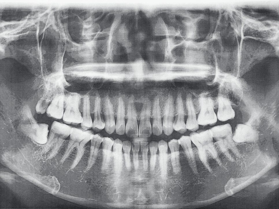
Magok-dong Dental Clinic_Panoramic
A panoramic image
shows not only the upper and lower teeth,
but also the gums and bone, wisdom teeth, and the jaw joint,
all of the oral cavity in one image.
When you first come to the clinic,
the majority of patients start with this image.
Why?
When you go to look at a house,
you do not check only one room first;
you look at the overall layout first.^^
A panoramic image plays exactly that role.
It gives you the feeling of,
“Oh, so this is what it looks like!”
like looking at a map of the mouth all at once.
That is why it is necessary in cases like these.
When you first visit the clinic,
when you want to see the overall condition at a glance,
when you want to check whether wisdom teeth are hidden,
or when you want to look for implants or major inflammation.
In short,
it is the basic image used to examine the entire mouth for the first time.
- Periapical Radiography
- A close-up image of a single tooth
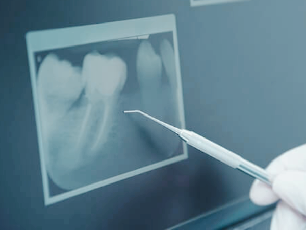
Magok-dong Dental Clinic_Periapical Exam
A periapical image
literally magnifies one tooth
so you can look at it very closely.
If a panoramic image is an “entire-mouth map,”
this is more like a magnifying glass
used to inspect one problem room up close.^^
Even if the tooth looks fine on the outside,
if there is inflammation inside
or decay affecting the nerve area,
this image makes it easier to check.
That is why it is taken in cases like these.
When one tooth keeps hurting,
when checking whether root canal treatment is needed,
or when inflammation at the tip of the tooth root is suspected.
It is a detailed image used to check whether everything is okay.
- Bite-Wing Radiography
- An image used to find cavities between teeth
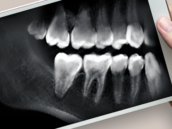
Magok-dong Dental Clinic_Bite-wing
A bite-wing image
shows the area between teeth,
the blind spots that are hard for a toothbrush to reach.
Even cavities that cannot be seen no matter how much you look in a mirror
are easier to check with this image.
If the outside looks fine,
but there is a sharp sensation only when chewing,
or food keeps getting stuck,
in many cases, this area is the cause.
So it is needed in situations like these.
When a small cavity is suspected,
or when looking for hidden cavities during a regular checkup.
In short,
it is an image that helps detect cavities between teeth that are not visible to the eye.
- Cephalometric Radiography
- An orthodontic image taken from the side of the face
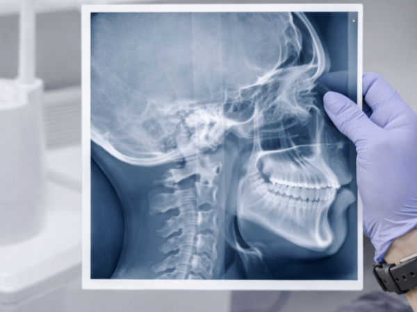
Magok-dong Dental Clinic_Cephalometric Radiography
A cephalometric image
is taken from the side of the face
to examine the balance of the teeth, jaw, and facial bones.
During orthodontic consultations,
you may have heard comments like,
“Your jaw is a little prominent,”
or “Your mouth looks like it protrudes forward.”
Rather than saying that based on intuition,
this image lets us view it objectively with numbers and angles.^^
So it is needed in cases like these.
Before orthodontic treatment,
when looking at jaw position or facial balance.
It is an orthodontic-specific image that shows the teeth and facial line together.
- 3-Dimensional Computed Tomography / 3D CT
- An examination that shows the mouth in three dimensions
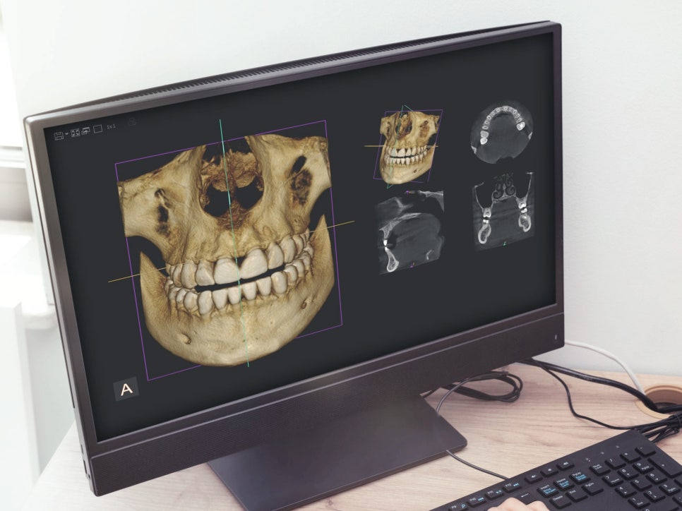
Magok-dong Dental Clinic_3-Dimensional Computed Tomography
Dental CT
is an image that lets you look at the teeth and jawbone in three dimensions.
If a regular X-ray is a flat map,
then CT is like a 3D navigation map.
You can turn it around and see the front, side, top, and bottom.
3D CT
combines images taken from multiple directions
to create a three-dimensional model of the mouth.
So what can you see?
The internal structure of the teeth,
the thickness and density of the jawbone,
and even where the nerves pass.
Even if things look fine on the outside,
it allows you to check in advance things like,
“How much space is there here?”
“Is the nerve too close?”
When placing implants, removing impacted wisdom teeth,
or before jawbone surgery,
CT can be helpful for understanding the structure more accurately.
Let’s summarize today’s content like this.
- Panoramic
→ A large map that shows the entire mouth at once
- Periapical image
→ A close-up image of one problematic tooth
- Bite-wing image
→ Finding cavities between teeth and hidden cavities
- Cephalometric radiography
→ An orthodontic image that shows facial and jaw balance
- 3D CT
→ A navigation map that lets you view the mouth in three dimensions

Magok-dong Seoul Dia Dental Clinic
At Magok-dong Seoul Dia Dental Clinic,
we value giving enough explanation before examinations.
“Why is this necessary?”
“Do I really have to take this?”
These are very good questions.
They are not unnecessary sensitivity;
they are questions from someone who truly cares about their teeth.^^
If you are confused about the types of examinations,
don’t search on your own—feel free to ask.
That one question can make treatment more accurate
and help you feel more at ease.
In the third part,
we will look at why dental radiographic exams are necessary.
Thank you for reading such a long post today.
[ This post was written in accordance with medical law for the purpose of providing accurate information about dental surgery and procedures. Infection and side effects may occur after surgery, so the procedure should be decided after sufficient consultation with a skilled medical professional. ]
[Magok-dong Seoul Dia Dental Clinic Dental Radiography Series]
-
Safety of dental radiographic examinations
-
Types of dental radiographic examinations
-
Reasons for dental radiographic examinations