Benign Skin Tumors: Ultrasound Diagnosis and
Clean Plastic-Surgical Excision
Hello.
I am Jun-ho Choi,
Director of Mutoel Plastic Surgery.
At Mutoel Plastic Surgery in Yongsan,
we provide care that considers both
our patients’ health and beauty
through close collaboration among
three specialists in dermatology,
plastic surgery, and pathology.

Many more people than expected
visit a clinic because of small lumps
or bumps beneath the skin that
began to feel soft or firm several
months earlier.
These lumps, which can occur in
various areas of the body—including
the face, neck, arms and legs, torso,
and scalp—are often mistaken for
ordinary acne or simple skin
troubles. As a result, people may
repeatedly press them with their
hands or try to squeeze them out.
However, forcefully attempting
extraction with one’s hands or
unsterilized instruments can cause
the internal sac to rupture, allowing
inflammation to spread into the
surrounding tissue.
This can lead to secondary bacterial
infection and severe inflammatory
reactions, causing the skin to become
red and swollen and potentially
resulting in extreme pain and other
side effects.
Most of these lesions formed beneath
the skin are benign skin tumors
located in the dermal or subcutaneous
fat layers.
Therefore, it is especially important
not to stimulate them arbitrarily, but
to first identify their exact cause
and location before proceeding with
systematic treatment.
Today, I will explain in detail the
precise ultrasound diagnosis and
plastic-surgical excision used to
safely and cleanly address “lumps
under the skin.”
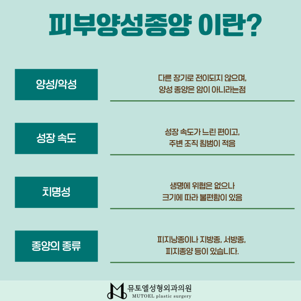
Representative Types and Characteristics of Benign Skin Tumors
Although benign tumors that occur
on the skin may look similar on the
surface, a detailed examination of
their interiors reveals considerable
differences in their causes, internal
structures, and contents.
The most representative condition is
an epidermoid cyst, also known as a
sebaceous cyst. It forms when the
opening of a sebaceous gland becomes
blocked, causing keratinous waste and
sebum to accumulate inside a cystic
sac.
An epidermoid cyst may show a small
central pore or opening. If forcibly
squeezed, it may release a foul-smelling
keratinous discharge or rupture
internally, developing into severe
inflammation.
In particular, an inflamed epidermoid
cyst may become strongly adhered to
the surrounding tissue, making
treatment very difficult with simple
excision alone.
If secondary cellulitis or a large
abscess develops, it may leave a
complicated scar after surgery.
Another representative condition is a
lipoma, a lump formed by excessive
proliferation of mature fat cells. It
feels soft beneath the skin and tends
to gradually increase in size over
time.
Other types of benign skin tumors may
also occur, including fibromas, which
result from the proliferation of
fibrous cells in the dermis, and
pilomatrixomas, which are observed as
hard, calcified lesions, particularly
in children and younger people.
Although these benign lesions are not
malignant tumors or cancer, they do
not disappear on their own. If left
untreated, they may continue to grow
and compress nearby nerves or blood
vessels.
They may also become visibly raised
and cause cosmetic concerns. For this
reason, it is advisable to receive an
accurate diagnosis early and plan
appropriate surgical treatment.
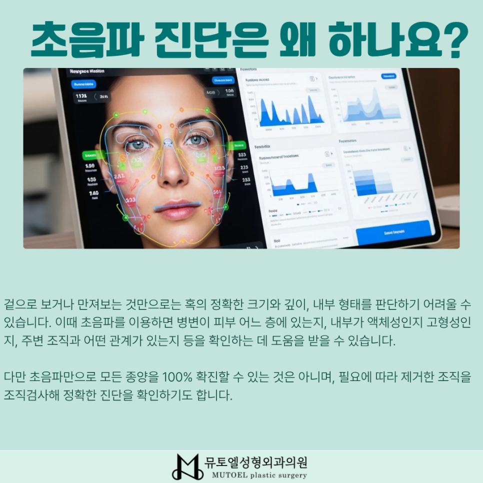
High-Resolution Preliminary Analysis Through Precise Ultrasound Diagnosis
The essential first step required for
successful plastic-surgical excision
is a precise preliminary evaluation
using high-resolution ultrasound.
Visual observation and palpation alone
have limitations when it comes to
clearly determining the exact
three-dimensional size, depth, and
boundaries of a lump located deep
beneath the skin in relation to the
surrounding tissue.
With the appropriate use of
ultrasound, it is possible to accurately
check whether the lesion extends
beyond the subcutaneous fat layer into
the fascia or muscle layer. This serves
as an important guide for carefully
determining the extent of dissection
during surgery.
This detailed preliminary examination
clearly establishes the boundaries of
the benign skin tumor and plays a
critical role in minimizing the extent
of the skin incision required during
surgery.
Real-time ultrasound also makes it
possible to check in advance whether
the lesion is in contact with small
nearby blood vessels or major nerves.
This helps substantially reduce the
risk of bleeding or nerve damage during
surgery while preserving normal tissue.
Sophisticated ultrasound diagnosis not
only maximizes surgical safety but
also provides the essential foundation
for minimizing postoperative scarring.
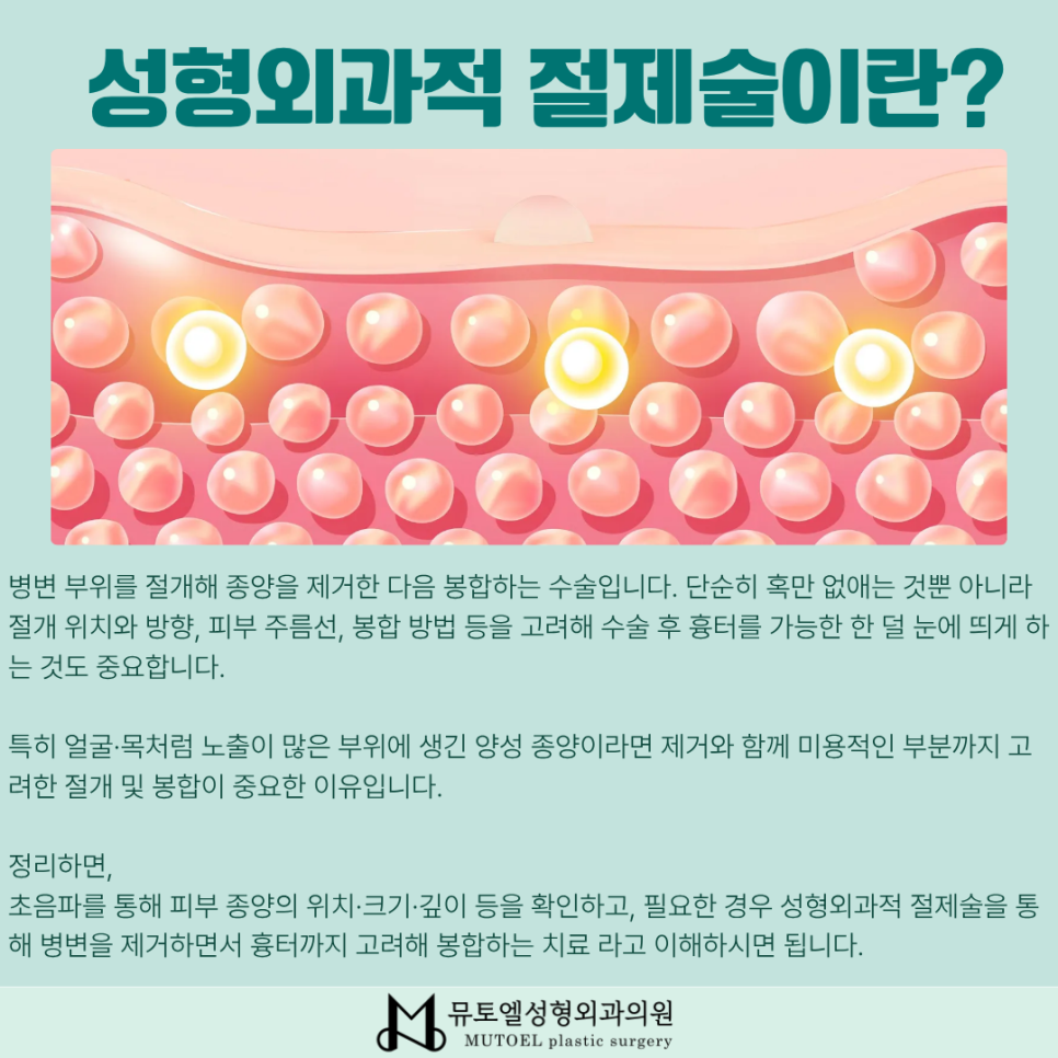
Plastic-Surgical Excision and Techniques for Minimizing Scars
To fundamentally treat a benign skin
tumor and prevent recurrence, it is
essential to completely remove the
entire sac surrounding the tumor, rather
than simply draining or squeezing out
its contents.
If even a small amount of the cyst wall
remains inside the skin, sebum may
accumulate again in the same area over
time, and the tumor may recur with a
high likelihood.
After the tumor has been completely
removed, a fine multilayer closure
technique is used, suturing the
subcutaneous tissue, dermis, and
epidermis in separate layers.
Through this delicate plastic-surgical
closure process, we minimize the skin
depressions and raised scars that may
be a concern after removal of a benign
skin tumor, helping create a smooth
skin surface.

Collaborative Care by Specialists in Dermatology, Plastic Surgery, and Pathology
One of the most distinctive features
of Mutoel Plastic Surgery Clinic is its
one-stop medical system, in which three
specialists in dermatology, plastic
surgery, and pathology work closely
together in one location.
All tissue safely removed through
plastic-surgical excision immediately
undergoes a detailed histopathological
examination by an in-house pathology
specialist.
This allows us to definitively confirm
whether the excised tumor is benign
and thoroughly assess the possibility
of any malignant transformation,
helping relieve our patients’ concerns.
During follow-up as the surgical wound
heals, a dermatologist provides
systematic, customized aftercare to
actively prevent and manage surgical
marks, pigmentation, and discoloration.
Depending on the postoperative stage,
scar-reducing laser or vascular laser
treatments may be performed as needed
to quickly reduce redness and help the
skin recover to a natural tone.
The precise excision and closure skills
of a plastic surgeon, the accurate
diagnosis of a pathology specialist,
and the attentive regenerative care of
a dermatologist come together in
harmony to provide the best possible
results for patients suffering from
benign skin tumors.
If you would like a more detailed
consultation about the surgery, please
feel free to contact us at any time.
We will always do our best for the
health and balanced beauty of each and
every patient.
This was Jun-ho Choi,
Director of Mutoel Plastic Surgery.
Thank you.