Hello, this is Dr. Jo Hyun-woo of Lip Plastic Surgery.
Today, I’d like to explain a possible complication after zygoma surgery—nonunion—using a real case.
As I consult with patients and follow up after surgery, I often meet many people who are worried about nonunion.
In zygoma surgery, nonunion means that the cut zygomatic bones have not fused together.
In fact, one thing many people overlook is that a more serious complication than sagging cheeks is nonunion.
As a result of nonunion, patients may experience not only sagging cheeks but also pain at the area where the bone has not fused, and they may feel the bone move when chewing.
Why does nonunion occur?
The causes of nonunion can include fixation problems, surgical issues, or patient carelessness.
It can happen if the fixation is not done properly, if no fixation is performed, or if a fixation pin breaks. In cases of an incorrect surgery, nonunion can occur regardless of whether fixation was done, so caution is necessary.
To prevent nonunion after zygoma surgery, you should limit strenuous chewing for about 6 weeks.
If the surgery was performed accurately, and you avoid excessively chewing tough or hard foods that put strain on the bone during the healing period, you can confirm by CT scan 6 weeks after surgery that the bone has fused well.
Therefore, if the surgery was done accurately, nonunion rarely occurs as long as you are careful during the healing period, and it can be sufficiently prevented through the medical team’s careful surgery and thorough postoperative care.
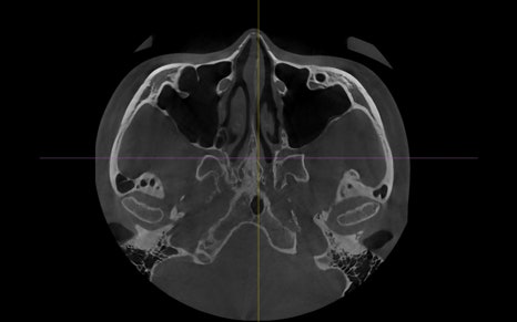
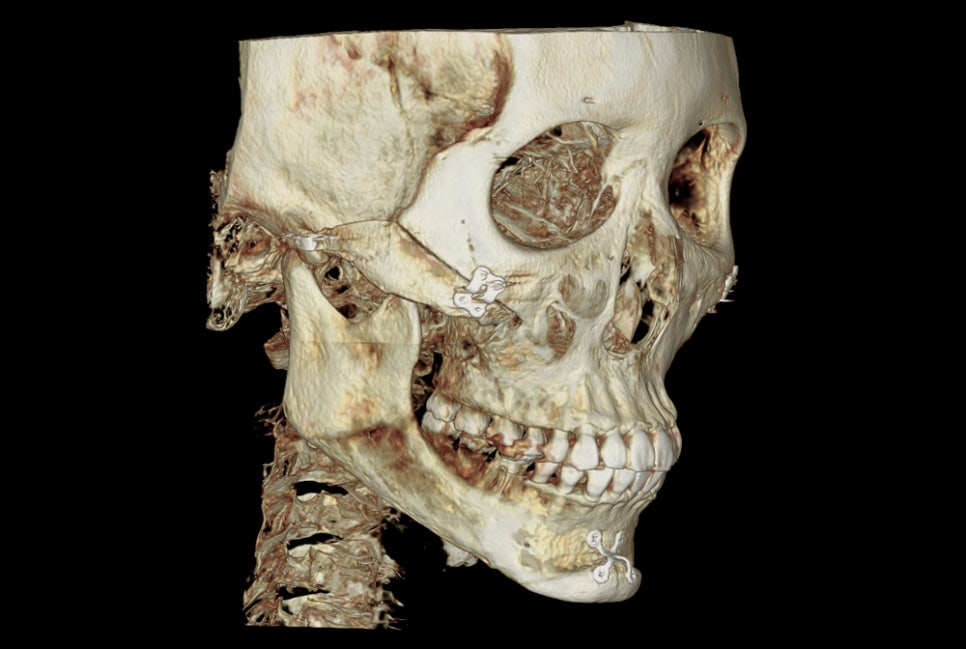
CT images showing the zygomatic bone has healed well without nonunion
This is where real patients often become curious.
Among those who come for consultations about revision zygoma surgery, many say, “I had a CT taken at another hospital, and it showed nonunion,” and visit us with that concern.
So when we take a CT scan,
They may say that the dark-looking area in the image means the bone has not fused, the surgery was done incorrectly, and revision surgery is needed.
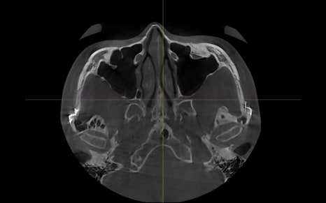
But if we adjust the shading slightly,
You can see that the bone has fused well.
A CT scan of the facial area that we usually take is created by overlaying multiple X-ray images taken at 5 mm intervals.
In fact, 5 mm is a very wide interval. CT scans taken at university hospital level are performed at 1 mm intervals.
However, the downside is that this takes a long time.
In conclusion, CT scans taken at 5 mm intervals to assess the overall shape can have a great deal of distortion.
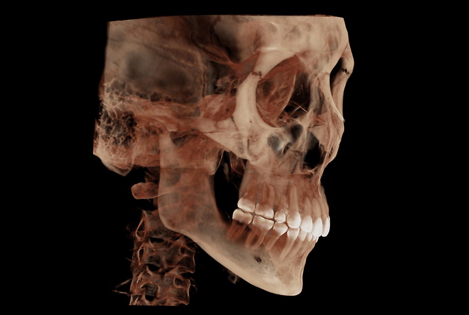
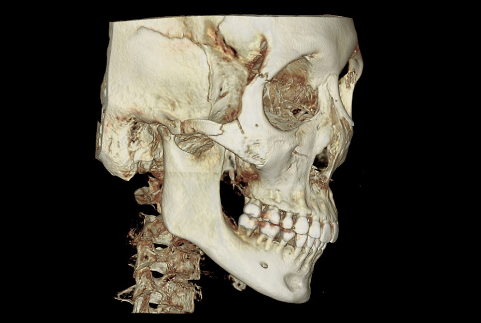
In addition, 3D CT is graphics created by a program based on the axial CT images taken in this way, so the distortion can become even greater.
Even if a 3D CT shows a hole in the bone or makes it look separated, when surgery is actually performed, there are many cases where the bone is completely fused.
In the following image, it looks as though there is a gap, but in reality there is no gap at all.
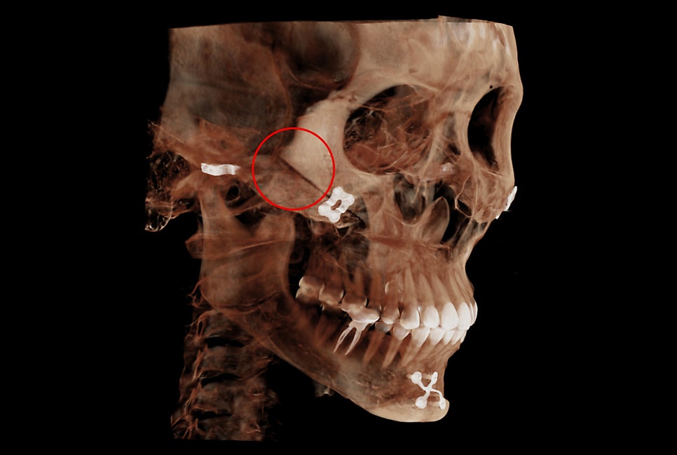
Looking at this, people may judge it as zygoma nonunion or a gap and worry.
This area has simply not yet undergone complete calcium deposition, and it is something that will eventually fill in over time.
Because zygoma gaps and nonunion can look slightly different depending on the viewing angle, an accurate diagnosis requires examining the entire CT from multiple angles.
There are also cases where a zygomatic bone that appeared to be nonunion later fuses again years after surgery, so rather than worrying too early at one month, three months, or even one year after surgery, it is important to stay relaxed and check the progress by taking CT scans at each stage after surgery.
For patients seeking consultation, it may be easier to understand if they think of bone images that look clearly separated as nonunion.
Some people may say that even a small separation means nonunion and that surgery must be redone, but if you keep in mind that these distortions exist, it may help you interpret the CT images.
Thank you.