Hello.
I’m 김하원 from 뷰티스닥터.
When bluish spots appear on the cheeks or temples,
many people initially think they are bruises or
melasma.
“Will it become lighter if I apply whitening cosmetics?”
“Can it also be managed with a standard pigmentation laser?”
These are questions we often hear in the consultation room.
With an Ota nevus, it is important to first examine
how deeply the pigmentation is located rather than focusing only on its visible color.
So today, I’ll summarize how to distinguish and evaluate it when managing an Ota nevus.
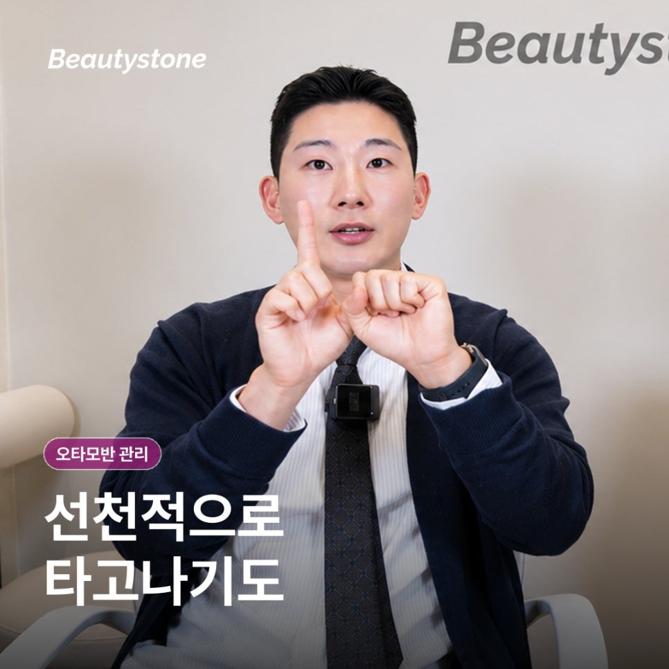
Three-line summary
- An Ota nevus is a lesion in which melanocytes are located
in the dermal layer,
so it requires a different approach from surface pigmentation.
- Treatment is adjusted based on the color and extent of the lesion
and the skin’s response, including the type, intensity, and interval of laser treatment.
- During recovery, avoid ultraviolet exposure and friction,
and monitor for strong reactions such as pain, blisters, or oozing.
- Why is an Ota nevus different from melasma or blemishes?
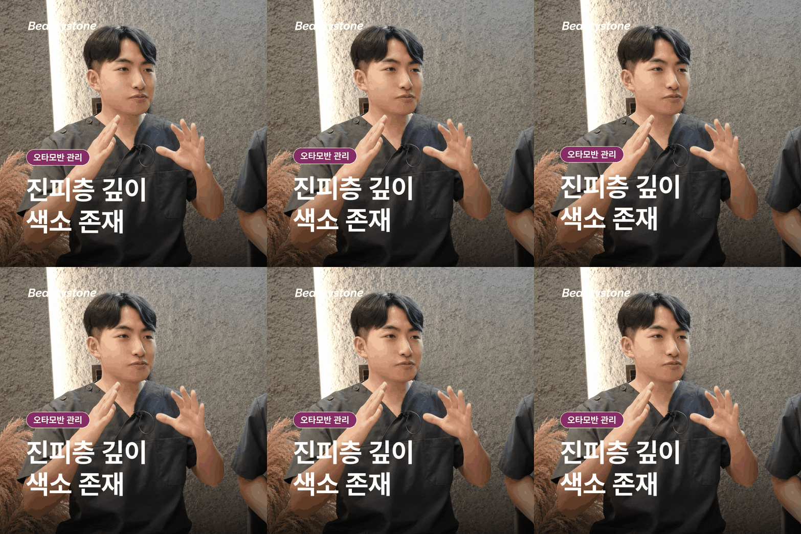
An Ota nevus is
a lesion that appears when melanocytes remain
in the dermis, the deeper layer of the skin.
The brown pigment located deep in the skin
can show through the skin and appear blue, blue-gray, or grayish brown.
It most commonly appears
on the forehead, temples, and around the eyes,
or on one side near the cheekbone.

However, not every grayish-brown spot that appears on both cheekbones
after adulthood
is the same type of lesion.
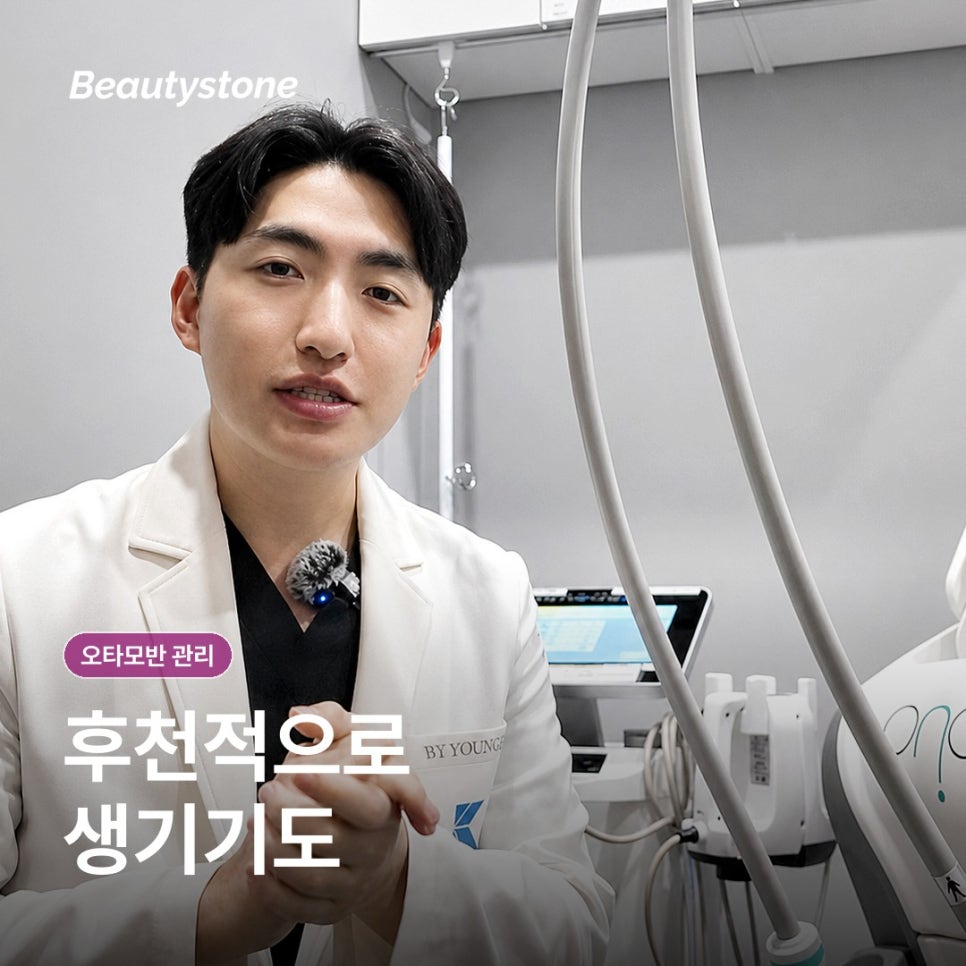
Acquired bilateral
nevus of Ota-like macules, melasma,
or post-inflammatory hyperpigmentation can also look similar,
so the timing of onset, distribution, and color tone should be evaluated together.
Even if it looks brown when covered with makeup,
the blue undertone may appear more prominent depending on the lighting or skin condition.
Rather than determining the lesion based only on a photograph,
also consider when it first appeared,
whether its area has expanded,
and whether it developed after inflammation or a procedure.
Therefore, the starting point for managing an Ota nevus
is not choosing a laser first,
but determining what type of lesion the current pigmentation represents.
- What factors determine how laser treatment differs?
When targeting pigment in the dermis,
a laser that selectively responds to pigment may be considered.
Even with the same Ota nevus, the wavelength, energy, and delivery method may vary
depending on how dark the color is,
whether the affected area is extensive,
and how close it is to the eyes.
Treating intensely in a single session
does not necessarily lead to a better outcome.
Excessive irritation may cause redness and blisters,
as well as post-inflammatory hyperpigmentation
or hypopigmentation.
Therefore, treatment is often divided into multiple sessions while monitoring the skin’s response,
and the number and interval of sessions
are determined based on the depth and extent of the lesion,
together with the skin tone and condition observed during consultation.

During the consultation,
rather than checking only the name of the equipment,
ask why that particular method was selected.
Immediately after treatment,
ask for an explanation of the expected redness and recovery process,

as well as the criteria that will be used to decide
when to proceed with the next session.
It is also helpful to check whether a fixed number of sessions is presented from the outset,
or whether the treatment plan is adjusted by comparing the skin’s response after each session.
- What information should be provided before the procedure?
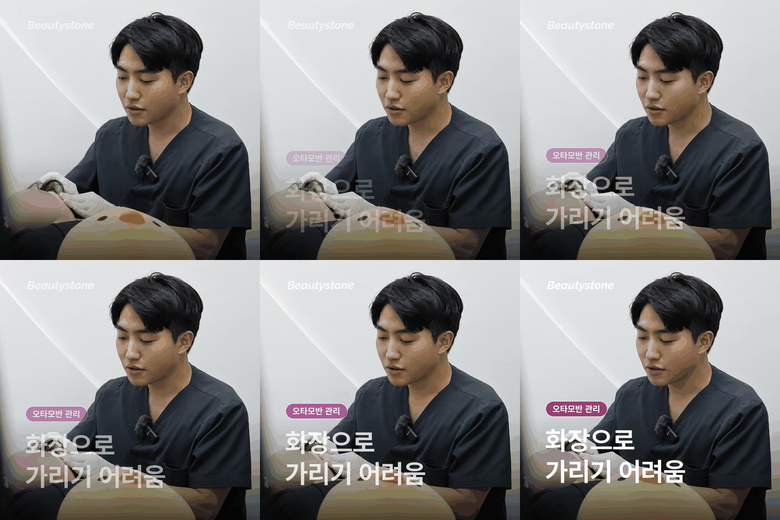
Medications you are currently taking and any allergies,
recent laser treatments or peels,
and a history of injectable procedures
are important information when establishing a treatment plan.
If you are taking medication that makes it difficult for bleeding to stop
or if marks tend to remain for a long time after wounds,
please inform the provider in advance.
If you have previously received pigmentation treatment,
it is helpful to explain which equipment was used and how many sessions were performed,
as well as how your skin responded afterward.
Knowing your previous response helps determine whether to repeat the same level of stimulation
or adjust the intensity or interval.
During the procedure, you may feel stinging and warmth,
as well as a sensation like a rubber band snapping against the skin.
It is helpful to receive an explanation before the procedure about the type of anesthesia,
protective equipment for the area around the eyes,
and how pain will be managed if it is severe.
- What should be monitored during recovery?

Immediately after the procedure,
redness and a burning sensation,
mild swelling or bruising, and thin scabs may appear.
Wash your face gently without rubbing,
and use any prescribed ointment or
moisturizer as directed.
Do not remove scabs by hand;
wait until they fall off naturally.
Even if the color does not become lighter immediately or temporarily
appears darker,
avoid trying to scrub away the skin flakes.
Applying multiple layers of whitening products or exfoliating products
may also increase irritation.
During recovery,
rather than trying to lighten the color quickly,
first focus on reducing friction and irritation
so that the skin barrier can stabilize.
When going outside,
apply sunscreen
and consider using a hat or parasol as well.
While redness and
warmth remain,
avoid activities that increase heat and friction, such as strenuous exercise, drinking alcohol, saunas, hot baths,
or facial massage.
If pain and swelling become increasingly severe,
or if large blisters, oozing, yellow discharge,
or spreading warmth develops,
do not dismiss it as ordinary
recovery; contact a medical institution.
If eye pain or vision changes occur after treatment around the eyes,
it is important to have them checked immediately.
Concluding remarks
Managing an Ota nevus
is difficult to view simply as a process of
removing deep pigmentation.
It should first be distinguished from similar pigmented lesions,
then continued by establishing a plan suited to the skin’s condition
and monitoring the recovery response.
Rather than judging based only on the visible color,
also consider when the lesion began and its distribution,
as well as the response to previous treatments.
In consultation,
it is helpful to ask specific questions not only about the diagnosis and treatment goals,
but also about the expected
skin response and the criteria for contacting the provider afterward.
Frequently asked questions
Q1. Can an Ota nevus be managed with cosmetics alone?
When the pigmentation is located in the dermal layer,
whitening cosmetics applied to the surface alone
may have limitations.
Q2. Is one laser treatment enough?
Because the depth and extent of the lesion vary,
it is difficult to determine in advance
the result of one treatment or the number of sessions needed.
Q3. Is a spot that appeared after adulthood an Ota nevus?
It may resemble
an acquired bilateral nevus of Ota-like macules
or melasma,
so the timing of onset, distribution, and color tone should be evaluated.
Q4. What symptoms should prompt contact after treatment?
Contact the provider if pain or swelling worsens,
or if large blisters, oozing, discharge,
or spreading warmth appears.
Thank you for reading this lengthy post today.
This has been 김가을 from 뷰티스닥터.