Hello, this is Dr. Seo Ho-yeon, the chief director of Garak Market Station Yonsei Dagam Dental Clinic.
Sometimes wisdom teeth do not erupt out of the gums and remain impacted.
So, when people come in for a checkup after a long time and are told that the tooth needs to be removed, they can feel quite startled.
Because they often hear from others that removing wisdom teeth is so painful... they start worrying even before treatment begins^^..
In particular, when they hear that the wisdom tooth is lying sideways or close to a nerve, they may feel afraid that it could be dangerous.
Reasons for Removing Wisdom Teeth
When the overall position is checked through a panorama, you can see that there are three wisdom teeth.
Do Wisdom Teeth Have to Be Removed?
If a wisdom tooth is properly aligned and functioning like a molar, there is no need to remove it.
However, in the example shown at the Garak Market Station dental clinic, the wisdom teeth are not fully serving the function of molars.
Above all, wisdom teeth are located at the very back, where it is difficult for a toothbrush to reach.
Especially if they are partially covered by the gums, food easily gets trapped, creating an environment where bacteria can multiply.
That is why cavities commonly develop in wisdom teeth.
Rather, if the condition is harmful to oral health and the tooth is not functioning,
it becomes something that can cause other problems as well, so in such cases, removal is advisable.
Horizontally Impacted Wisdom Tooth
The two wisdom teeth in the lower jaw still seem to be growing straight,
but for some reason, the one wisdom tooth in the upper jaw may look slightly strange in direction.
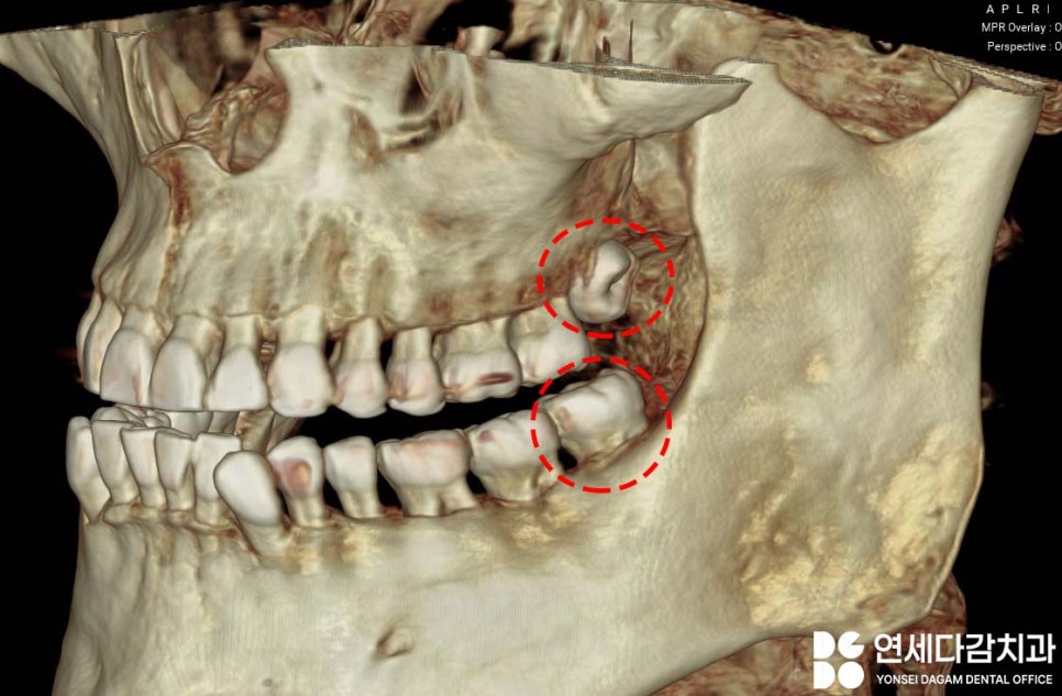
Unlike the lower jaw, you can see that the top surface of the upper wisdom tooth is facing toward the cheek side.
When the three-dimensional structure is analyzed with CT,
it is possible to precisely determine the direction in which the tooth is growing,
how close it is to surrounding structures,
and the shape of the roots as well.
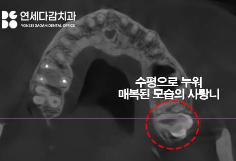
In this case, it is lying horizontally
and is partially impacted.
A horizontally impacted wisdom tooth refers to a tooth lying on its side and buried in the gums or bone.
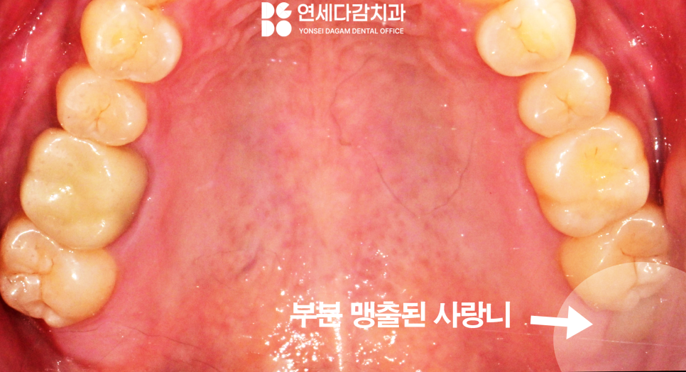
Sometimes it is partially erupted,
but impacted cases or partially erupted cases are more difficult to remove than normally erupted wisdom teeth.
Moreover, if it is close to anatomical structures,
extra caution is needed.
Why Treatment Analysis Is Important
For example, at the Garak Market Station dental clinic,
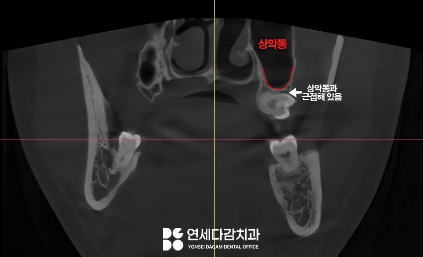
there are times when a tooth is close to the maxillary sinus.
The maxillary sinus is an empty space inside the upper jawbone.
If the wisdom tooth root is close to this space, caution is needed during extraction.
It is covered by a mucous membrane,
and if the wisdom tooth root is close to the maxillary sinus,
this membrane can tear during extraction.
If the membrane is perforated,
the mouth and the maxillary sinus become connected,
and food or liquid may come out through the nose,
with a risk of sinusitis developing.
Therefore, through CT analysis,
the distance to the maxillary sinus is checked in advance,
and a treatment plan is established to prevent perforation.
In the lower jaw,
attention must be paid to damage to the inferior alveolar nerve.
The inferior alveolar nerve canal is a passageway inside the lower jawbone that contains a nerve.
If the wisdom tooth root is close to this nerve canal,
the nerve may be compressed or damaged during extraction.
If the nerve is damaged,
it is characterized by altered sensation in the lower lip or chin.
There may be tingling
or reduced sensation,
and in most cases it recovers over time,
but in rare cases it may last for a long time.
Therefore, CT is used to check the distance to the nerve canal and the positional relationship between the roots and the nerve canal,
and the plan is made in a way that minimizes the risk of nerve damage.
Removing it without causing damage
requires a high level of skill.
At the Garak Market Station dental clinic, such anatomical relationships are analyzed
in order to establish a safe treatment plan.
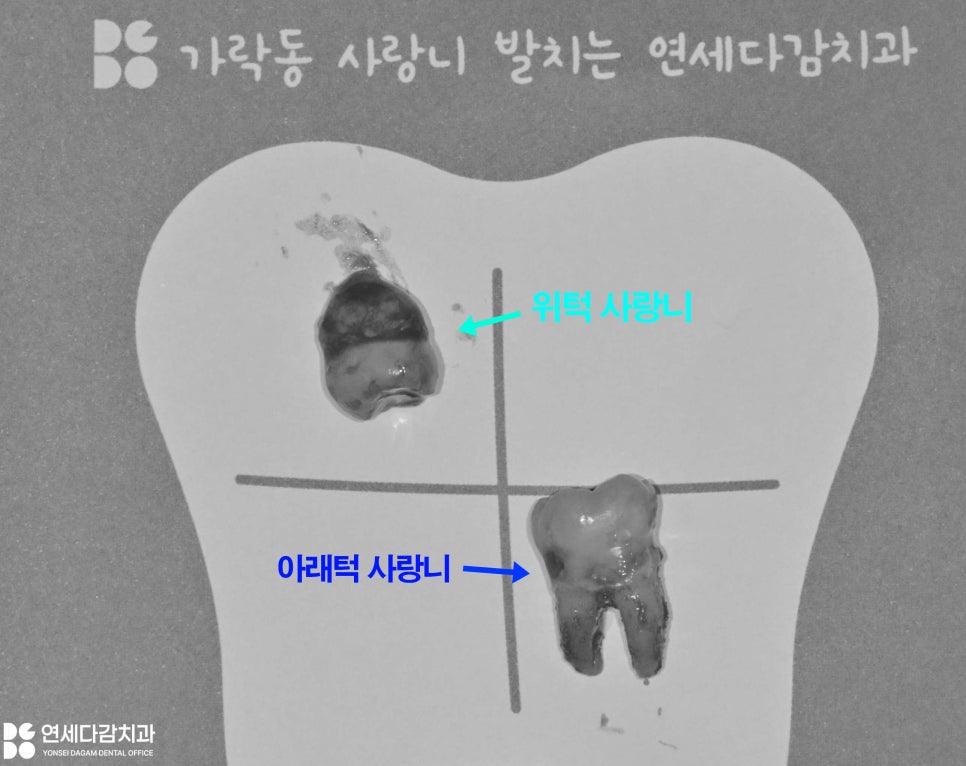
A horizontally impacted upper wisdom tooth
is removed by making an incision in the gum.
Since an impacted tooth is covered by the gum,
it must be exposed through an incision before it can be removed.
In this case, fortunately,
it was possible to remove the tooth whole
without dividing it.
Compared with what was expected on CT,
the root shape was simple,
so it could be removed without sectioning,
and no maxillary sinus perforation occurred.
Suturing was performed to help with hemostasis,
completing the extraction.
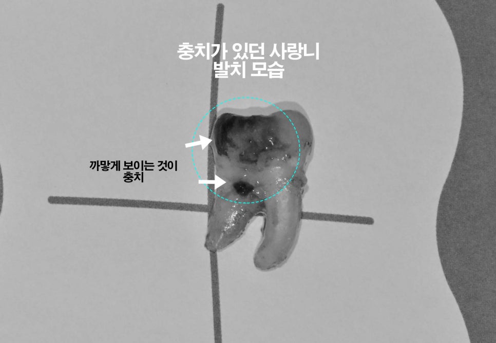
Below, you can see that after removal, the tooth was blackened
and decay had progressed.
For teeth like this, removal is recommended^^
As shown today at the Garak Market Station dental clinic,
when you hear that a wisdom tooth is lying sideways or close to a nerve,
you may worry.
However, through CT analysis,
if the tooth position, root shape,
and relationship with surrounding structures are accurately identified,
even a horizontally impacted wisdom tooth can be safely extracted.
If you are dealing with a similar problem,
we recommend getting an accurate examination from an experienced specialist
and having it removed while you are still as young as possible.
If you are usually very afraid of dental treatment,
there is also a method using sedation anesthesia (conscious sedation).
We hope you can regain your oral health through a treatment that suits you,
and we will come back next time with useful information.
This was Garak Market Station Yonsei Dagam Dental Clinic!