Hello, this is Ceramic Dental Clinic. Today, we’re going to take a look at root resorption, one of the representative side effects of orthodontic treatment.
During orthodontic treatment, root resorption, in which the roots of the teeth become shorter, occurs very slightly in almost all patients, but severe resorption that becomes a problem is reported in the literature at around 1–5%. Because the numbers vary widely depending on the study population and measurement method, it is safer to understand them as a “range” rather than an absolute value.
What is root resorption, and why does it occur during orthodontic treatment?
Root resorption is the phenomenon in which the apical portion of a tooth root is subtly shortened during orthodontic treatment. You can think of it as the root embedded in the jawbone gradually dissolving from the end as it is exposed to the continuous force applied by orthodontic appliances.
To use an analogy, it is similar to a tree root planted in the ground whose tip is slightly worn down due to changes in the surrounding soil. Up to a certain point, the human body can recover on its own, but once the shortening goes beyond a certain level,
it remains as an irreversible change.
When orthodontic force—that is, the force from wires or appliances pushing the teeth—stimulates the tissues around the root, odontoclasts become active and resorb the root tip. They are similar in form to osteoclasts, which resorb bone, but the difference is that their target is the hard tissue of the tooth. Since this is a physiological response that occurs to some extent in all orthodontic patients, the real clinical question is not whether it happens, but how far it has progressed.
How often does root resorption occur? Incidence and severity
Mild root resorption is observed in most orthodontic patients, but severe resorption that can affect function has been reported in some literature at around 1–5%. Because measurement methods and follow-up periods differ from study to study, it is better to treat these numbers as reference values.
Severity is usually divided as follows. This is a simplified version of criteria commonly used in clinical practice, such as the Malmgren classification, and the cut-offs vary slightly depending on the society or textbook.
| Grade | Degree of resorption | Clinical significance |
|---|
| Mild | Shortening of less than 1 mm | No effect on tooth function |
| Moderate | Shortening of 1–4 mm | Requires careful observation |
| Severe | Shortening of 4 mm or more, or more than one-third of the root | May affect tooth prognosis |
It occurs more often in the front teeth, especially the upper front teeth. That is because the roots are relatively short and the amount of movement during orthodontic treatment is large. Molars have multiple, thicker roots, so the frequency of resorption is relatively lower.
It has been reported that even severe root resorption can often be detected early enough to preserve the tooth.
However, this does not apply uniformly to every case.
Am I in a high-risk group? Risk factors that increase root resorption
Risk factors are broadly divided into tooth and patient factors, and treatment factors. Even if one side is strong, it does not immediately lead to severe resorption, but the risk accumulates as the two axes overlap.
Tooth and patient factors
-
Short or pointed roots. There is less room to begin with, making them more vulnerable to resorption.
-
Curved roots (root curvature). Force tends to concentrate at the root tip during movement.
-
Family history. If there is a history of root resorption after orthodontic treatment in the family, closer attention is needed.
-
Systemic disease. Conditions that affect bone metabolism, such as thyroid dysfunction, may change the risk level.
-
Teeth that retained primary teeth for a long time. Because the developmental process was irregular, the root shape may also be altered.
Treatment factors
-
The longer the treatment period, the greater the cumulative exposure, and thus the cumulative risk.
-
Excessive orthodontic force. Strong force increases tissue damage before it promotes faster movement.
-
Vertical movement. The risk is especially high with intrusion (movement that pushes a tooth into the bone).
-
The greater the amount of movement, the more the root tip is exposed.
Having multiple risk factors does not necessarily mean severe resorption will occur.
However, if you know about them in advance before treatment, you can adjust the frequency of monitoring.
Step-by-step guide to root resorption management from before treatment to after completion
Management is not a one-time test at a single point in time, but a continuous process that runs from before treatment through the retainer phase.
Step 1. Detailed examination before treatment
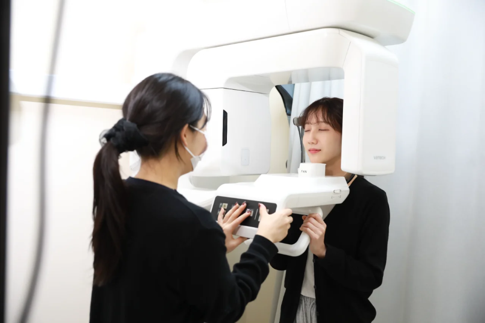
A panoramic X-ray (a flat image that captures the entire dentition on one film) is used first to check root length and shape. If curvature or signs of resorption are suspected, CBCT (three-dimensional CT) is used to examine the bend and direction in three dimensions.
Step 2. Risk disclosure when making the treatment plan
If high-risk factors are identified, there is a process of hearing about the risks and alternatives before treatment begins. This is because even for the same person, the level of risk varies depending on which teeth are moved and by how much.
Step 3. Regular monitoring during treatment
In general, a mid-treatment X-ray is taken 6–9 months after orthodontic treatment begins to check for changes in the roots (the recommended timing varies depending on the clinician and the society). If abnormal findings are seen, the orthodontic force may be reduced or the appliance may be paused for 1–2 months.
Step 4. Response when severe resorption is found
If the root has shortened by 4 mm or more, or if more than one-third of it has been lost, the treatment direction itself is reconsidered. Adjusting the target position or ending treatment early may also be reviewed. It is helpful to remember that this is not the result of poor treatment, but a choice appropriate to the situation.
Step 5. Care after treatment is completed
Even after orthodontic treatment is finished, it is safer to check the root condition with X-rays once every 1–2 years. For teeth with moderate or greater resorption, the interval is often shortened further.
Procedures patients often overlook and common misconceptions to correct
Misconception 1: “If root resorption occurs, the tooth will soon fall out.”
Even with severe resorption, the tooth does not immediately become lost. Even if the root is shortened, daily chewing function is often not significantly affected if the surrounding jawbone supports it well enough. However, because the long-term lifespan of the tooth may be reduced, it is better to step up the level of daily care.
Misconception 2: “If it hurts, that means the resorption is severe.”
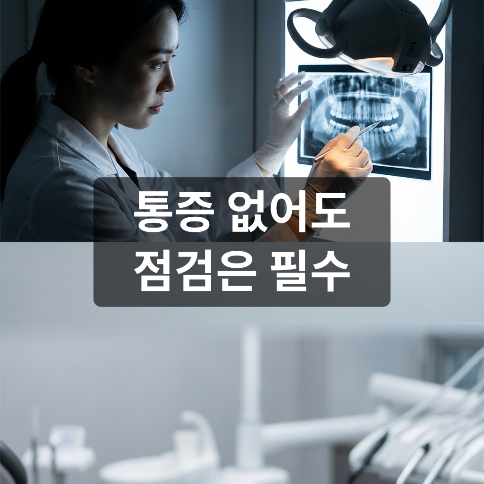
Root resorption is usually not painful. Because it progresses with almost no symptoms, regular X-rays are practically the only way to detect it early. Rather than feeling reassured because there is no pain, it is safer not to skip the scheduled mid-treatment checkups.
Misconception 3: “Clear aligners do not cause root resorption.”
Whether it is brackets (metal appliances) or clear aligners, the biological principle of moving teeth is the same. The risk is influenced much more by movement direction, distance, and duration than by the type of appliance.
X-rays during treatment may feel inconvenient,
but they are the fastest way to detect resorption that progresses without pain.
Orthodontic root resorption: frequently asked questions
Q1. If root resorption occurs, do I have to stop orthodontic treatment no matter what?
Not necessarily. If the resorption is mild, treatment is usually continued. If it is moderate or greater, the orthodontic force may be reduced or treatment may be temporarily paused and then reassessed, and in severe cases the treatment goals themselves may be adjusted. Rather than “stop no matter what,” a step-by-step response is closer to the standard.
Q2. Can shortened tooth roots grow back?
Once a root has shortened, it does not regenerate. However, if orthodontic force is stopped, additional resorption often stops there. The earlier it is detected, the more root length can be preserved.
Q3. Does adult orthodontic treatment carry a higher risk of root resorption than adolescent treatment?
Age itself is less of a variable than treatment duration and amount of movement. In adults, tooth movement tends to be slower and treatment can take longer, so the time exposed to risk may increase as a result.
Q4. Does root resorption concentrate in specific teeth only?
It is most common in the upper front teeth. That is because the roots are shorter and the movement distance is large in that area. It can also occur in the lower front teeth or molars, but the frequency is relatively lower.
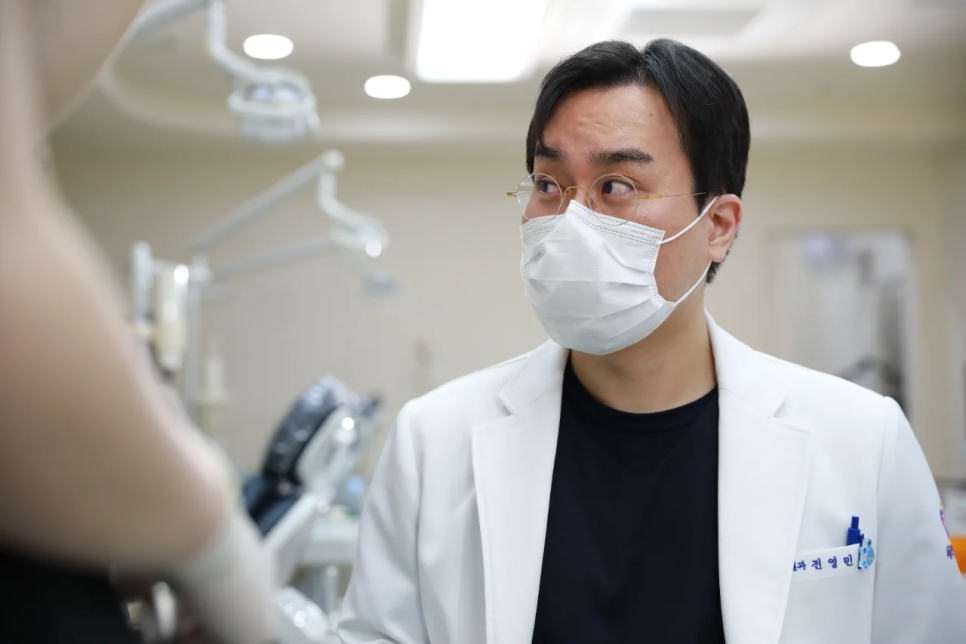
How to check the root resorption management system when choosing an orthodontic clinic
When choosing an orthodontic clinic, it is easier to compare if you look first at the monitoring system rather than the procedure price or advertising copy. Root resorption is a change that progresses without pain, so in the end, “how often and in what way it is checked” determines tooth preservation.
It may be helpful to look into the following points.
-
Whether CBCT is used when necessary in addition to the pre-treatment panoramic X-ray
-
How the timing of mid-treatment checkup X-rays is explained during treatment
-
Whether the response procedure for root resorption is explained in advance
It is difficult to reduce the incidence of root resorption itself to zero, but if you know the risk factors in advance and do not skip checkups, you can lower the proportion that leads to problematic amounts of resorption.
Because orthodontic treatment requires a significant investment of time and money, you should look into the details and prepare thoroughly, but with enough attention, cases where root resorption becomes a problem are rare. If you have been postponing orthodontic treatment because you were worried about root resorption, how about getting an examination and consultation this time?