Sometimes people bring in a mirror photo and ask whether surgery is necessary because their face looks very crooked. But when they actually take a front-facing photo, there are often no noticeable differences. On the other hand, there are also cases where real functional problems with chewing or speech are present.
For facial asymmetry, the key to deciding the treatment direction is not simply whether it exists, but what the cause is, how severe it is, and whether functional problems are accompanied. Depending on the type of cause, non-surgical treatment may be enough in some cases, while orthognathic surgery may be needed in others. In this article, we’ll go through those criteria and options one by one.
Facial asymmetry: how much is considered a level that “needs treatment”?
No face is perfectly symmetrical from left to right. According to facial measurement studies, even in adult faces, differences of about 2–3 mm are often observed somewhere on the left and right sides, and this is considered within the physiological range, meaning normal.
The clinically relevant criteria are a bit more specific.
If the facial midline is deviated by 3 mm or more,
if the occlusal plane (the surface where the upper and lower teeth meet) is tilted,
or if functional problems with chewing or speech are present,
then a professional evaluation is needed.
There is one helpful point to keep in mind. The face we see in the mirror is a left-right reversed image. Because of that, people often feel asymmetry more strongly than it actually is.
When compared with a front-facing photo, it is common for there to be a fairly large difference between how you perceive yourself and how others actually see you.
If there are no functional problems and only you are bothered by it, there is a separate step to look at before surgery.
Different causes mean different treatments: first comes type-specific diagnosis

Facial asymmetry can broadly be divided into three types depending on the cause. Without correctly identifying the type, it is difficult to expect the best result from any treatment.
The differences are easy to see at a glance in the table below.
| Type | Main cause | Representative condition |
|---|
| Skeletal | Asymmetry of the bones themselves | Mandibular condylar hyperplasia, hemifacial microsomia |
| Muscular | Imbalance in muscle volume and tension | Masseter hypertrophy (overdevelopment of one side of the jaw muscle) |
| Soft tissue | Differences in soft tissue such as skin and fat | Hemifacial atrophy |
Diagnosis starts at the first visit with visual measurement, facial photos, and dental arch examination. Then, if needed, additional detailed tests are performed in sequence, such as panoramic X-rays (X-rays of all teeth and jawbones), cephalometric X-rays (lateral skull X-rays), 3D CBCT (three-dimensional CT), bone scans, and MRI. Which tool is used depends on the suspected cause, and not all tests are done from the start.
This is especially important for minors, because bone growth may still be ongoing. Since the timing of surgery can only be determined after growth has completely stopped, bone age testing or cephalometric comparison at regular intervals must come before the decision for surgery. If this check is skipped, asymmetry may progress again after surgery due to further growth.
Can it be improved without surgery? A complete guide to non-surgical treatment
Non-surgical treatment can have supportive effects for muscular and soft tissue asymmetry. However, it is important to understand from the beginning that the scope of application for skeletal (bony) asymmetry is limited.
Botulinum toxin (for masseter hypertrophy): When one jaw muscle is overdeveloped, botulinum toxin is injected to reduce muscle volume. In the literature, the duration of effect is reported to average about 4–6 months, but there are individual differences depending on treatment history and muscle response, so repeat treatments are needed to maintain the effect. Pregnancy, breastfeeding, and neuromuscular diseases are major contraindications, and repeated treatments may change the pattern of effectiveness over time, so keeping a record of treatment history is helpful.
Hyaluronic acid filler (soft tissue volume correction): This method adds volume to the hollow side to create visual balance. Rather than solving the underlying cause, it is a way to correct the appearance. If injected into a blood vessel, serious complications such as vascular occlusion or skin necrosis can occur, so choosing an experienced practitioner is important.
Orthodontic treatment is a key tool for precisely addressing dental asymmetry.
If there is a large skeletal discrepancy as well, the pre- and post-surgical orthodontic stages become a major factor that determines the stability of the result,
so treatment is planned together within a cooperative care system involving orthodontics and oral and maxillofacial surgery.
When temporomandibular joint disorders or postural imbalance are the cause, splint therapy (a dental protection device) or physical therapy may be applied first. This is because correcting only the appearance without resolving the cause easily leads to recurrence.
If surgery is necessary: the orthognathic surgery process and the reality of recovery
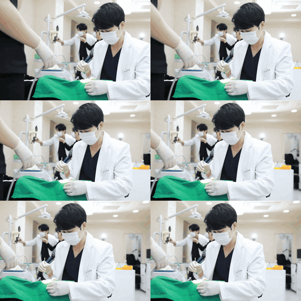
If surgery is decided on for skeletal asymmetry, you should first understand that the entire treatment schedule is a long journey lasting 2 to 4 years. The process is broadly divided into four stages.
-
Pre-surgical orthodontics (1–2 years): This is the stage in which the position of the teeth is adjusted in advance to suit the surgery. This stage cannot be omitted because it determines the stability of the bite after surgery.
-
Orthognathic surgery: Performed under general anesthesia and carried out within a cooperative system between orthodontics and oral and maxillofacial surgery.
-
Post-surgical orthodontics (6 months–1 year): This stage completes the remaining tooth alignment after surgery. Only after this final orthodontic step is finished does the final bite settle into place.
-
Retainers: Worn after orthodontic treatment is completed to maintain the results.
After surgery, hospitalization usually lasts 5–7 days, and intermaxillary fixation (a procedure that binds the upper and lower teeth together to keep them fixed) is maintained for about 6–8 weeks. During the intermaxillary fixation period, it is difficult to open the mouth sufficiently, so a liquid diet (such as thin rice porridge, porridge, and blended foods) is maintained during the same period. Swelling usually begins to noticeably decrease around 3 months after surgery, and it takes 6 months to 1 year to fully confirm the final result.
Common misconceptions patients repeat: knowing them in advance can prevent regret

These are misconceptions often heard in facial asymmetry treatment. If decisions are built on the wrong assumptions, they can easily lead to unnecessary costs or disappointment with the results.
"If I just get orthodontic treatment, my facial asymmetry will improve too."
Orthodontic treatment is a procedure that produces precise effects in the area of tooth alignment. If a large skeletal discrepancy is also present, the pre- and post-surgical orthodontic stages become a key factor that determines the stability of surgical results, so it is important at the diagnostic stage to determine whether orthodontics alone is enough or whether treatment should be carried out together within a cooperative care system.
"Botox and fillers can also correct skeletal asymmetry."
These are tools that deal with muscles and soft tissue in a supportive way, so they can help visually soften asymmetry, but they do not change the position of the bones themselves.
"Isn’t double jaw surgery just a cosmetic operation?"
It is a highly complex surgery performed under general anesthesia, and when the pre- and post-operative orthodontic periods are included, the treatment process takes 2 to 4 years. It is completely different in nature from a simple cosmetic procedure, so sufficient cooperative consultation is recommended before making a decision.
"Oriental medicine clinics, massage, or exercise devices can fix skeletal asymmetry."
It is helpful to review your options while keeping in mind that the clinical evidence for skeletal asymmetry is currently limited.
Before deciding which option is right for me, what should I do first?
Depending on whether the cause of the asymmetry is skeletal, muscular, or soft tissue, the treatment direction changes completely.
Whether functional problems such as chewing or speech difficulties are present, and whether the midline deviation exceeds 3 mm, are concrete starting points for determining whether treatment is needed.
If the asymmetry is only minor and noticed by you alone, it may help to first examine the perception itself before deciding on treatment.
It is much safer to use the information in medical advertisements only as general reference and rely on the results from imaging tests and professional consultations.
In a system where orthodontics, oral and maxillofacial surgery, and plastic surgery work together, it is a good idea to visit a nearby dental clinic or oral and maxillofacial surgery clinic as the first consultation point and receive basic imaging tests first. Confirming where the asymmetry originates is the fastest starting point for every treatment decision.