Among facial contouring surgeries, square jaw surgery, which is performed to soften the outline of a sharp lower face, has now become a very common procedure.
However, some people return to the clinic years after square jaw surgery because they are concerned that bone may have formed again at the operated angle area.
Based on my more than 20 years of experience, I would like to explain how the bone at the surgical site changes over time after square jaw surgery.
The material I am sharing here is part of what I submitted for publication in an international plastic surgery journal.
I will leave out the scientific and academic details and post only simple, easy-to-understand content.
After square jaw surgery, as time passes, the bone at the surgical site undergoes a remodeling process that follows a slightly different pattern from the immediate postoperative state.
Of course, these changes can vary greatly from person to person.
This difference is because the action of the muscles covering the bone varies from person to person.
Let’s look at the first case.
This was a patient with very well-developed masticatory muscles covering the mandible, and the CT images show the state 1 year and 3 years after square jaw surgery, as well as 1 year after a second procedure to refine some regrown angle bone.
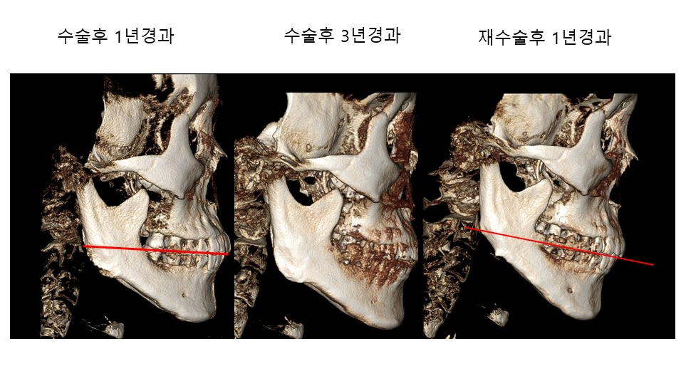
The condition of the right lower jaw bone 1 year and 3 years after the first surgery, and 1 year after the second surgery
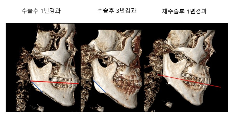
The blue line marked in the CT image above is the line I refined during surgery.
One year after surgery, it shows a state in which part of the angle area is forming, and after 3 years, a fairly developed state can be seen.
To refine the regrown bone at the angle, I smoothed the angle bone along the line marked in the second image.
The condition of the left lower jaw bone 1 year and 3 years after the first surgery, and 1 year after the second surgery
Let’s look at the second case.
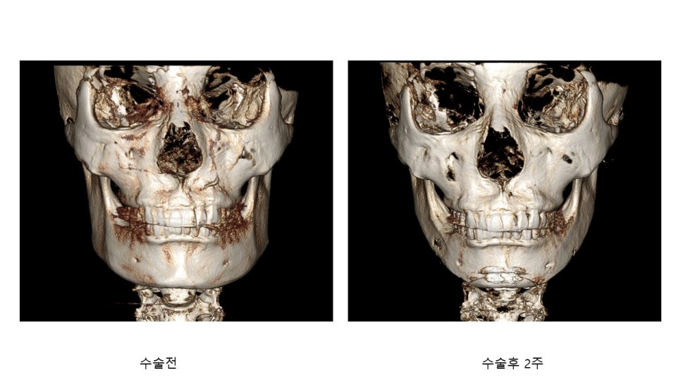
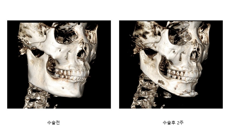
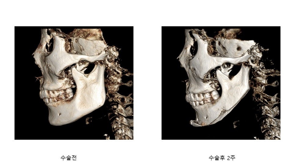
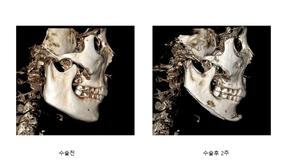
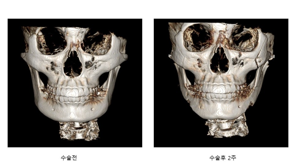
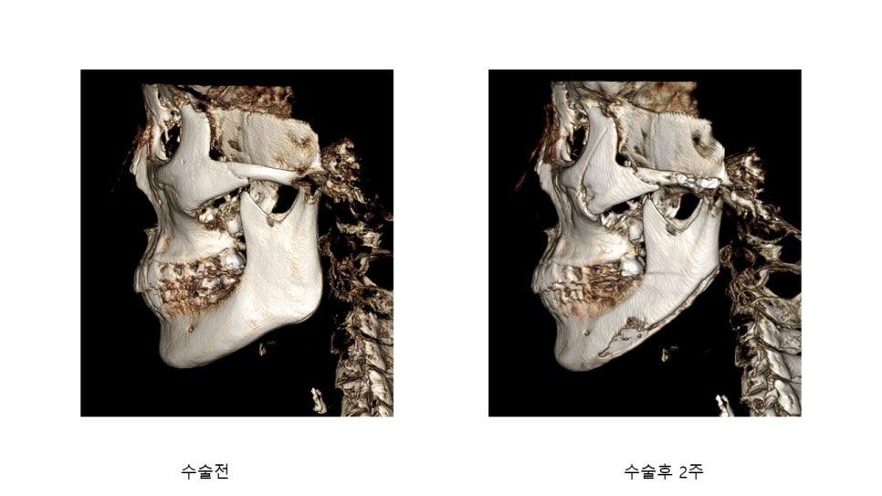
CT images before square jaw surgery including chin advancement and 2 weeks after surgery
Comparing the CT images before surgery and 2 weeks after surgery, the cortical bone of the mandibular body, which had the thickest cortical bone, has decreased significantly, making part of the bone appear as if it were hollowed out.
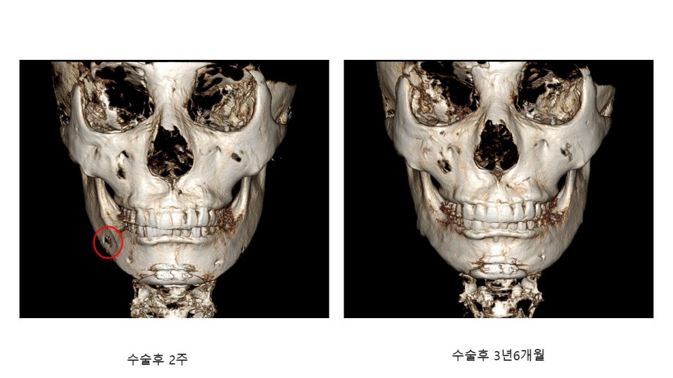
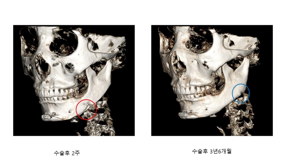
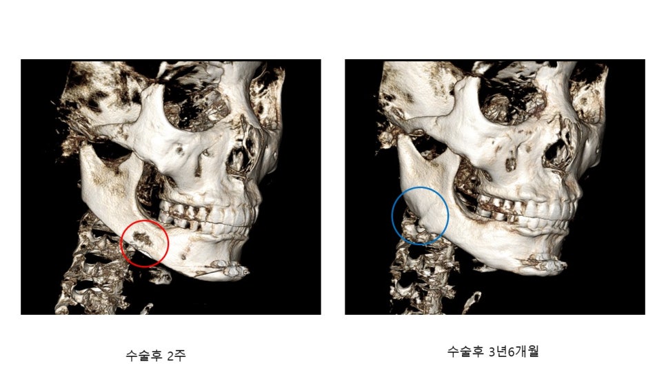
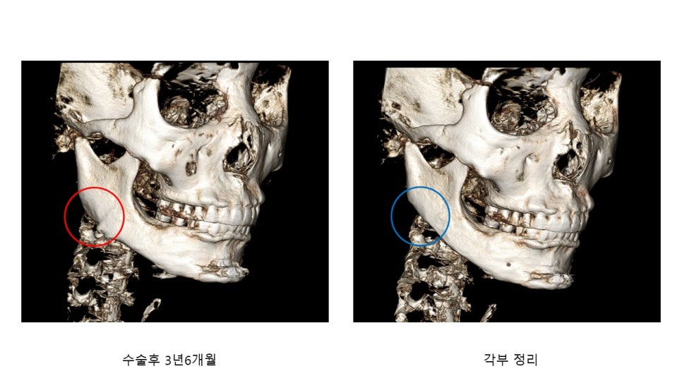
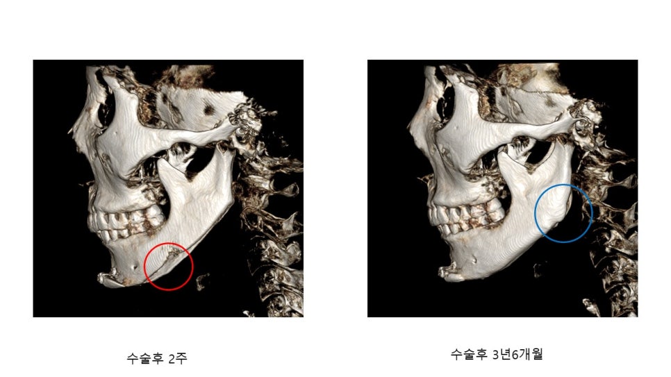
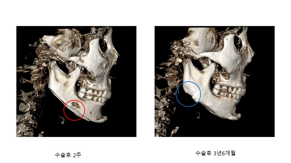
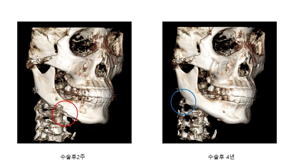
Comparison CT images 2 weeks after surgery and 3 years and 6 months after surgery
At 3 years and 6 months after surgery, the mandibular body (red circle), which had appeared somewhat hollowed out at 2 weeks after surgery, is covered with white cortical bone, and some bone has formed at the angle on the right side (left side on the screen).
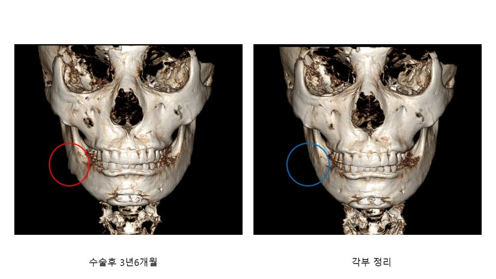
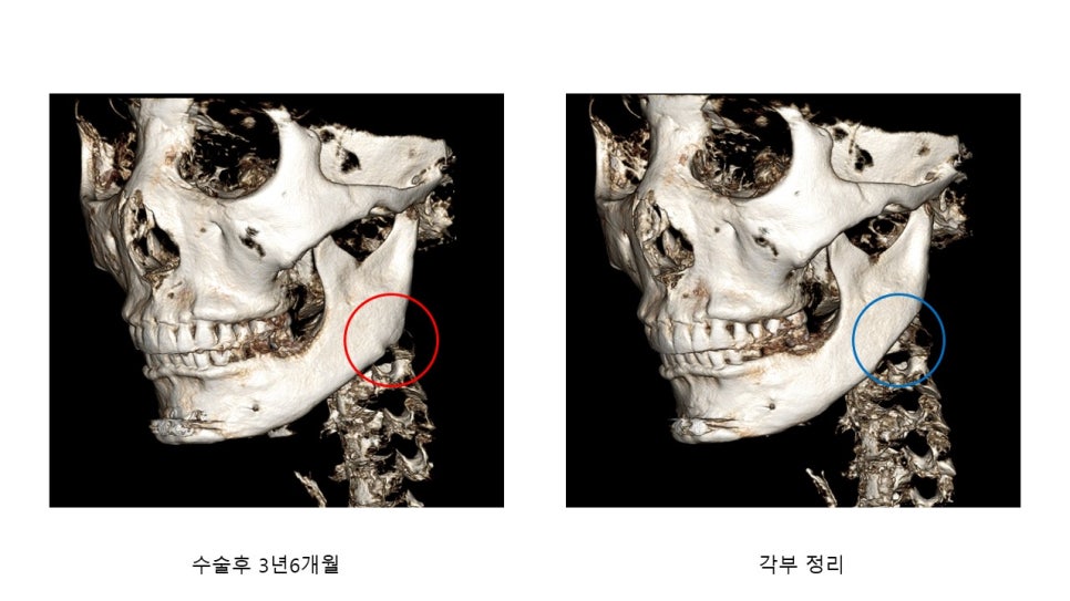
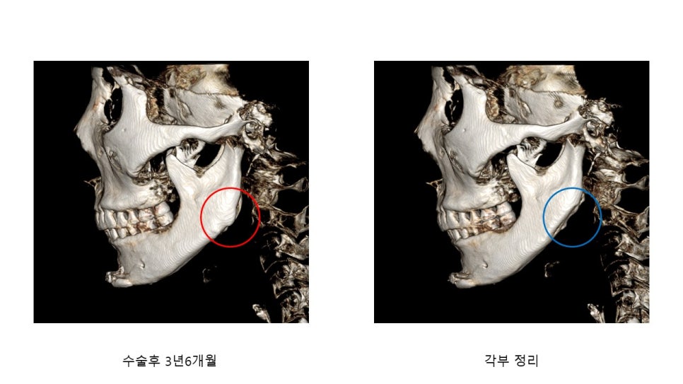
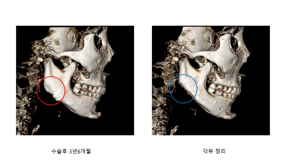
CT after the patient returned 3 years and 6 months after surgery and requested that the newly formed angle be refined again
This is the CT after refining the angle again at the patient’s request, after they returned 3 years and 6 months after surgery because they wanted the newly formed angle to be smoothed again.
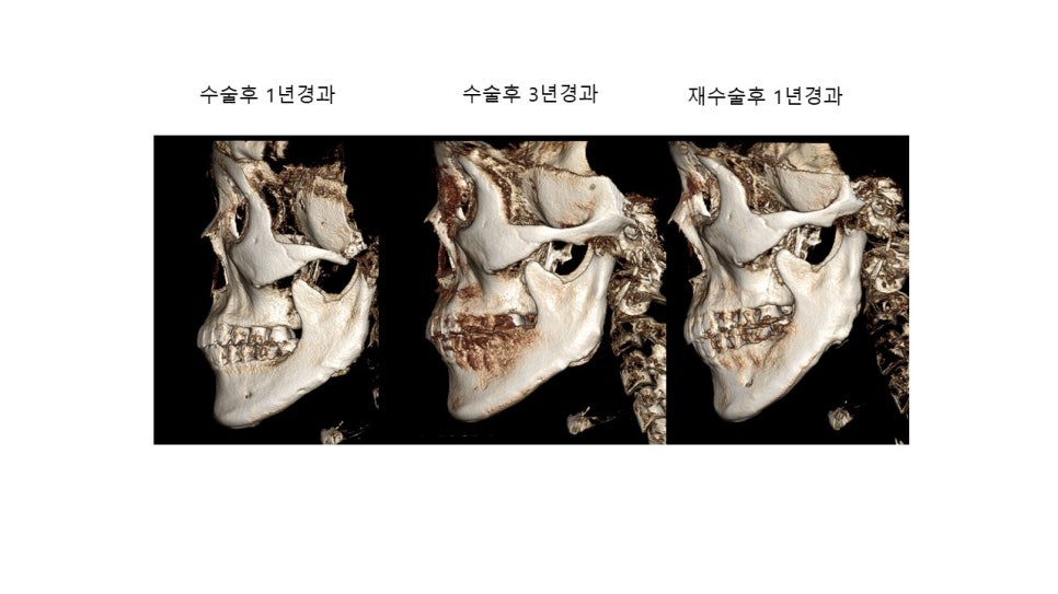
Let’s look at the third case.
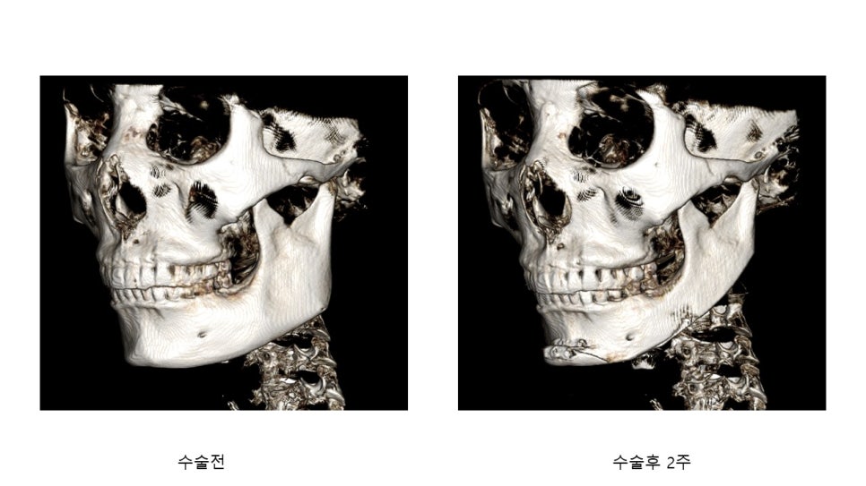
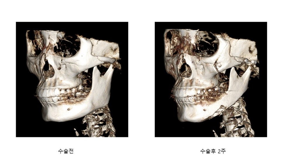
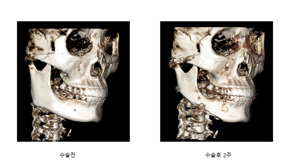
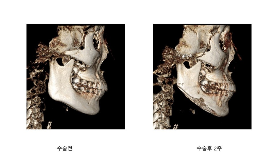
CT images before surgery and 2 weeks after surgery
Because the outline of the mandibular body before surgery appeared somewhat strong, the surgery was performed with the goal of reducing as much of the body bone as possible, and the CT at 2 weeks shows that the body bone appears somewhat hollowed out.
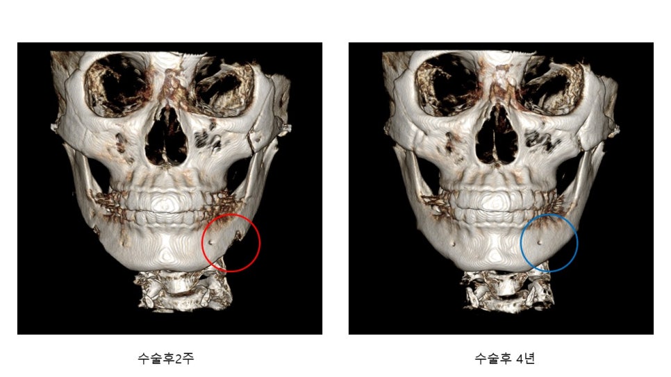
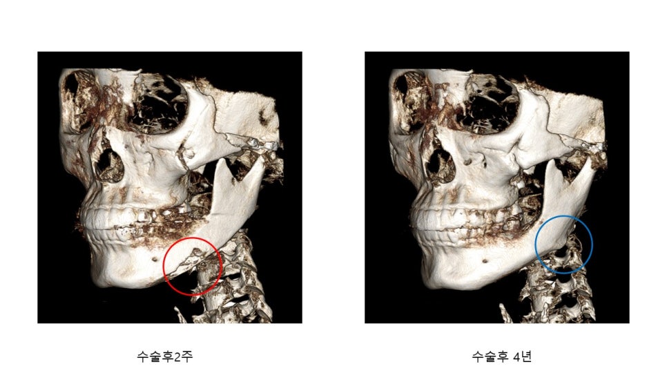
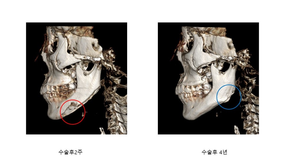
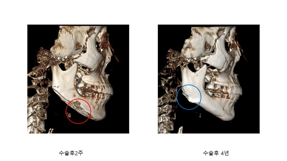
CT images 2 weeks after surgery and 4 years after surgery
If you look at the CT images 2 weeks after surgery and after 4 years, you can see that the bone in the area that appeared hollowed out is covered with cortical bone.
The cases I have introduced are very diverse, but since the content would become too long, I will conclude by introducing them only briefly.
In actual practice, after square jaw surgery, significant regrowth of bone at the angle does not always occur.
It can be said that cases in which the angle significantly forms again are less common than cases in which it does not.
However, the areas that appear hollowed out in the early postoperative period after reducing cortical bone can be observed to be covered again with white cortical bone even after just a few months.