During zygoma reduction surgery, is a non-fixation technique used?
The zygoma is a fundamental structure that forms the contour of the central part of the face. Because of its three-dimensional shape, it can be conveniently divided into the body of the zygomatic bone, which determines the contour of the front cheek or 45-degree cheek, and the zygomatic arch, which determines the contour of the side cheek. (Figure 1)
This zygoma is connected to the maxilla in the front and the temporal bone in the back, and the masseter muscle pulls downward on the lower side. Revisional zygoma surgery
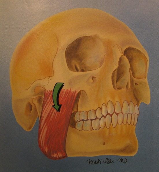
(Figure 2)
In surgery to reduce the shape of the zygoma, reducing the prominence of the front cheekbone is relatively simple and can be done sufficiently through an intraoral approach alone.
However, in order to effectively and sufficiently reduce the side cheek, the zygomatic arch, which widens as it extends backward, must be brought inward to achieve a satisfactory side-cheek reduction effect. (Figure 3) Revisional zygoma surgery
In such surgery, to reduce the width of the side cheek, the two points of the zygomatic arch that form the contour of the side cheek must be cut and fixed in a new position.
The reasons are as follows. Revisional zygoma surgery
First, a clear osteotomy is necessary in order to bring the zygomatic complex (the zygomatic body and zygomatic arch) inward.
Second, if fixation is not performed after a proper osteotomy, the zygomatic complex will, due to the action of the masseter muscle pulling the zygomatic arch downward, not only fail to unite properly (nonunion) but also sag downward. Revisional zygoma surgery
Third, the authors (Yang Du-byeong and Jeong Jae-yeong) previously published a method of intraosseous osteotomy using a green-stick fracture twice in the PRS Journal. Dr. Yang Du-byeong has originality in this method, which induces a bent shape like a bamboo stem without completely cutting the anterior osteotomy site.
However, the posterior part is completely osteotomized, and in very rare cases where only a small reduction of the side cheek is needed, it is possible to leave it unfixed and position the bones so that they interlock with each other.
But to bring it sufficiently inward, it is essential to adequately move the completely osteotomized posterior portion inward and then fix it.
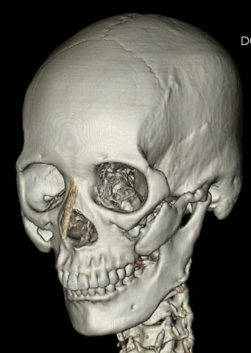
In addition, when there are questions or concerns about the results after zygoma surgery, it is essential to perform a three-dimensional CT scan and accurately check the postoperative shape of the zygoma.
The 3D-CT image below shows an image in which the front part of the zygoma was not fixed, creating a gap and causing it to sag downward.
The above content was presented by the author at the "2005 Autumn Conference of the Korean Society of Plastic and Reconstructive Surgeons" and the "2008 Symposium of the Council of Private Practice Plastic Surgeons".
http://www.lavianps.com
bach1@nate.com