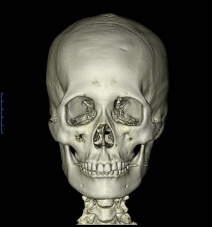
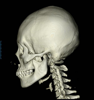
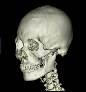
Because the facial bones are surrounded by soft tissue, it is not easy to estimate their three-dimensional shape.
In such cases, if the facial area is scanned at precise, regular intervals and then reconstructed by integrating according to signal intensity, the resulting three-dimensional image can be observed from any desired angle.
Reconstructing tomographic images in three dimensions allows not only the facial bones, but also to some extent the soft tissues of the face, to be reconstructed and viewed.
This kind of three-dimensional tomography (3DCT, 3D-CT) can be very useful in facial contouring surgery.
In particular, for ordinary people who do not have medical knowledge, it is almost impossible to understand their own facial bones from plain X-ray images alone.
Therefore, if you want to directly check the changes in your facial bones before and after facial contouring surgery, or if you are considering revision surgery because you are dissatisfied with the results after facial contouring surgery, it may be helpful to confirm the appearance of these facial bones through three-dimensional tomography.
http://www.lavianps.com
bach1@nate.com