Among the facial bones that determine the face shape, the zygomatic bone not only determines the contour of the middle part of the face, but also plays the biggest role in determining the width of the face.
As public understanding of facial contouring surgery has broadened, there is also a concern that the triggers and standards for deciding on facial contouring have become overly relaxed.
In the opinion of the author, who has performed facial contouring surgery every day for more than 10 years, even an experienced board-certified plastic surgeon needs to invest at least 1 hour and 30 minutes of operating time to properly perform zygoma reduction surgery.
The reasons are as follows.
First, because the zygomatic bone is a structure surrounded by facial muscles, subcutaneous fat, and skin, unpleasant results from inaccurate zygoma reduction surgery become more severe over time and as a person ages.
Even if the surgery is not done properly, it may not be noticeable for 1 to 2 years after surgery.
Second, if zygoma reduction surgery was performed incorrectly and secondary corrective surgery must be considered, the operation can be much more difficult than in areas that are easier for the surgeon to access.
Third, the zygomatic bone has a bridge-like structure connecting the anterior maxilla to the posterior temporal bone, and because the masseter muscle pulls downward, if the bone is cut and not properly fixed, it can lead over time to the bone sagging downward or parts of the bone being resorbed and disappearing.
Looking at the results of inaccurate zygoma reduction surgery, they are as follows.
1. When the zygomatic bone was cut and left without proper fixation, causing the zygomatic bone to drop downward
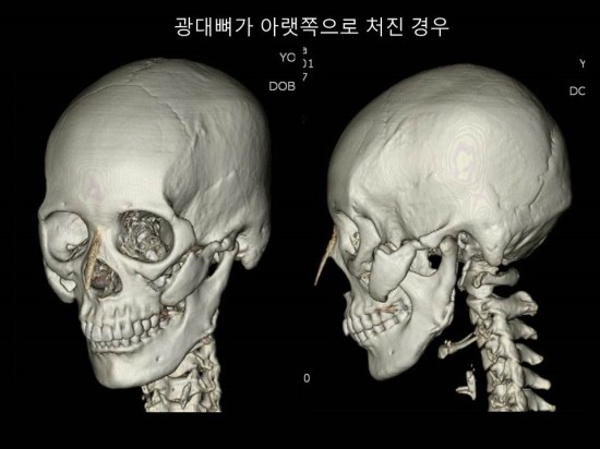
As can be seen on the 3D CT of the patient above, if the front part of the zygomatic bone is sufficiently cut but left without fixation, the zygomatic bone sags downward and a large gap forms between the bones.
In such cases, although zygoma reduction surgery was performed, the face may only look saggy, creating a result that makes the person look older and more depressed.
The reason is that the masseter muscle pulls the zygomatic bone downward, so if it is left unfixed, the bone sags downward over time, as shown above.
It may not be noticeable for several months or even up to a year after surgery, but as time passes, wrinkles deepen through the gap between the front bones.
2. When only the front zygoma was cut and forced into fixation, making the face look wider toward the back
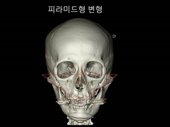
The 3D CT above shows a case in which only the front zygoma was cut and the side zygoma was not properly osteotomized, yet fixation was forced.
Because the front part was fixed, the zygomatic bone did not sag downward, but due to a pyramid-shaped deformity that widens from front to back, it created an optical illusion that the face looks wider toward the back after surgery.
In such cases, zygoma revision surgery is relatively easy.
3. When the zygomatic bone was cut appropriately and then forcibly pushed in by hand
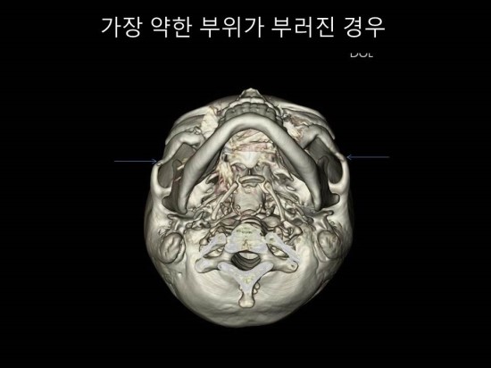
The 3D CT above shows a case caused by pressing the zygomatic bone down by hand without accurately cutting the front and back portions.
In such cases, even if the surgery can be completed within 30 minutes, the weakest middle part of the zygomatic arch breaks, producing a step-off deformity.
In these cases, zygoma revision surgery becomes the most dangerous and difficult.
Therefore, zygoma reduction surgery should be decided carefully, and it is necessary to accurately analyze changes in the appearance of one’s bones before and after surgery through 3D CT.