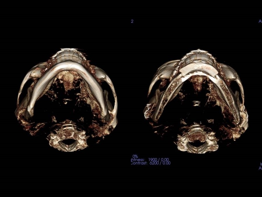
After cheekbone reduction surgery, the continuity of the area connecting the front cheekbone to the side cheekbone is the most important.
When analyzing cases in which people come to see me because of unsatisfactory results after cheekbone reduction surgery using various methods, the following phenomena can be observed.
- Loss of continuity in the front cheekbone
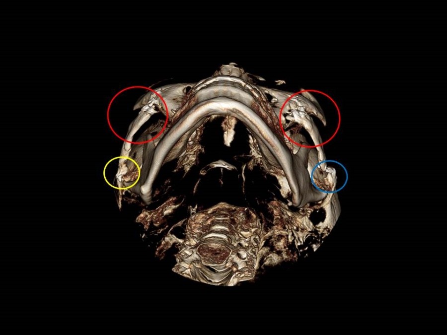
If you look at the inner area of the red circle in the 3DCT above, you can see that the continuity of the area connecting the front cheekbone to the side cheekbone has been disrupted, causing a step deformity.
To examine the continuity of the area connecting the front cheekbone to the side cheekbone, it is important to check the 3DCT from various angles while viewing the cheekbone arch from below the jaw. In such cases, there may be no obvious sign early after surgery because of swelling, but as time passes, step deformities and hollowing may occur in the area connecting the outer corner of the eye to the temple.
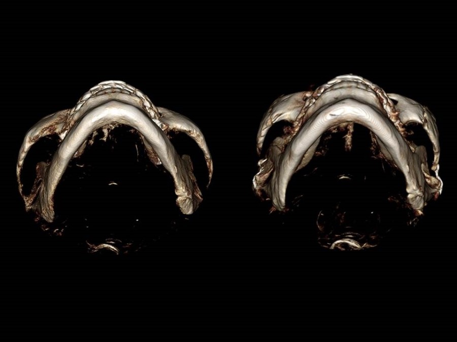
- Excessive hollowing of the front cheekbone
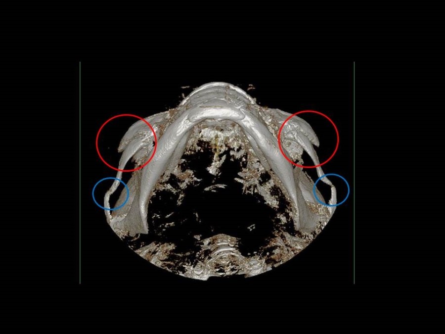
In the 3DCT above, you can see the step deformity in the cheekbone area inside the red circle and the posterior cheekbone arch with inaccurate osteotomy and fixation not properly done (simply bent while the bone was broken).
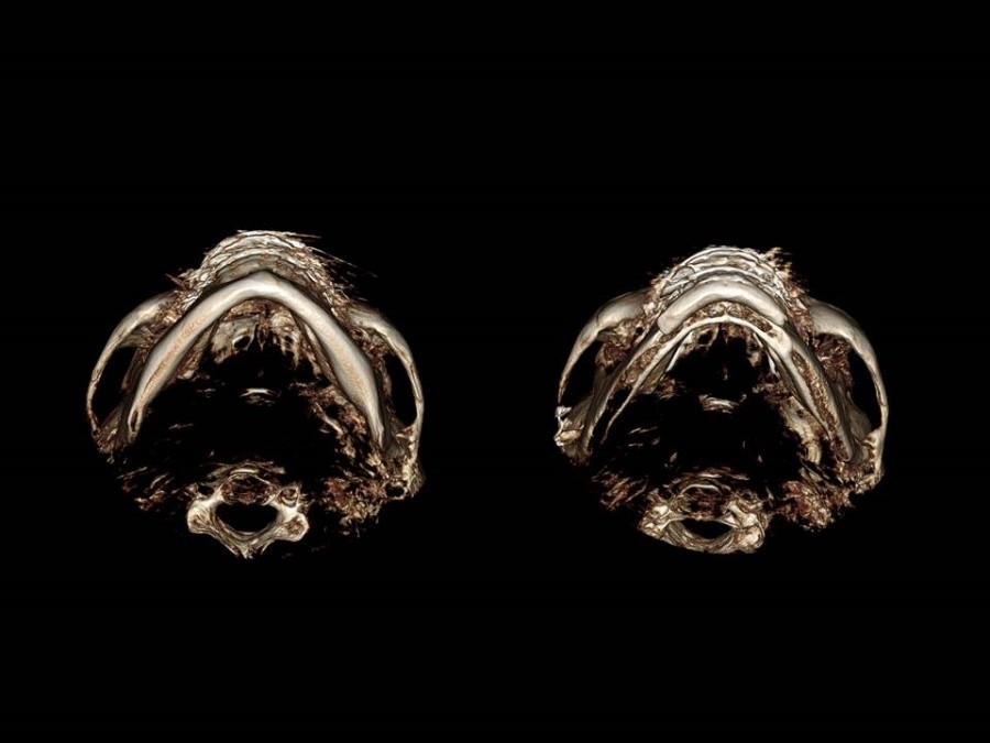
- Incorrect osteotomy position at the side cheekbone osteotomy site
In the 3DCT above, the posterior osteotomy position of the cheekbone arch to reduce the side cheekbone is not appropriate.
If osteotomy is performed in this area, not only will there be no reduction effect on the side cheekbone, but a step deformity will occur at the osteotomy position within the blue line, and the broad area in front of the sideburns may not decrease at all.
In such cases, revision surgery can be extremely difficult.
Next,
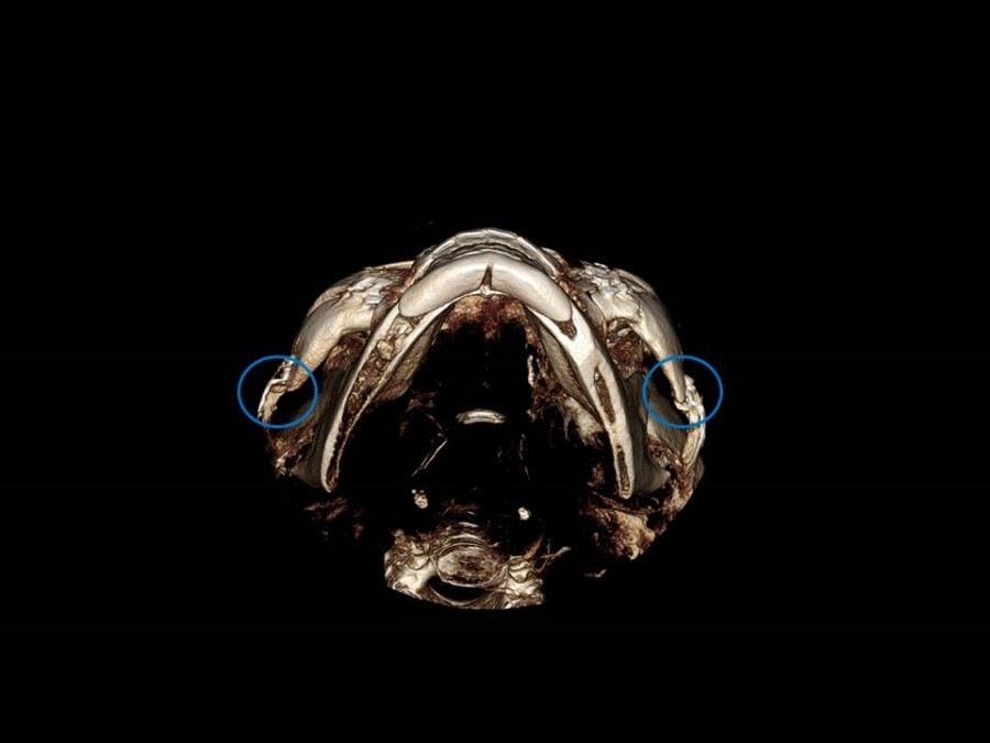
the following are cases in which there is no reduction effect on the side cheekbone at all after cheekbone reduction surgery.
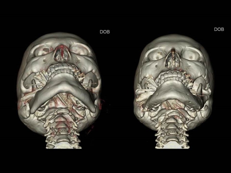
- Cheekbone reduction surgery through a scalp incision
The 3DCT above shows a case in which cheekbone reduction surgery was performed through a scalp incision, but there was no change in the posterior osteotomy position of the cheekbone inside the blue circle, so there was no reduction effect on the side cheekbone at all (no reduction in facial width).
If the width of the cheekbone arch, which becomes wider toward the back, is not effectively reduced through a scalp incision while reducing the front cheekbone area, there is a risk that the face will appear flat from the front and wider toward the back.
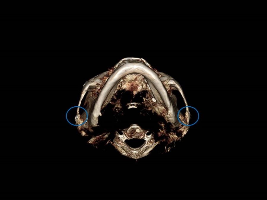
-
Incorrect selection of the posterior osteotomy position of the cheekbone arch
-
Inaccurate fixation of the cheekbone arch and posterior osteotomy site
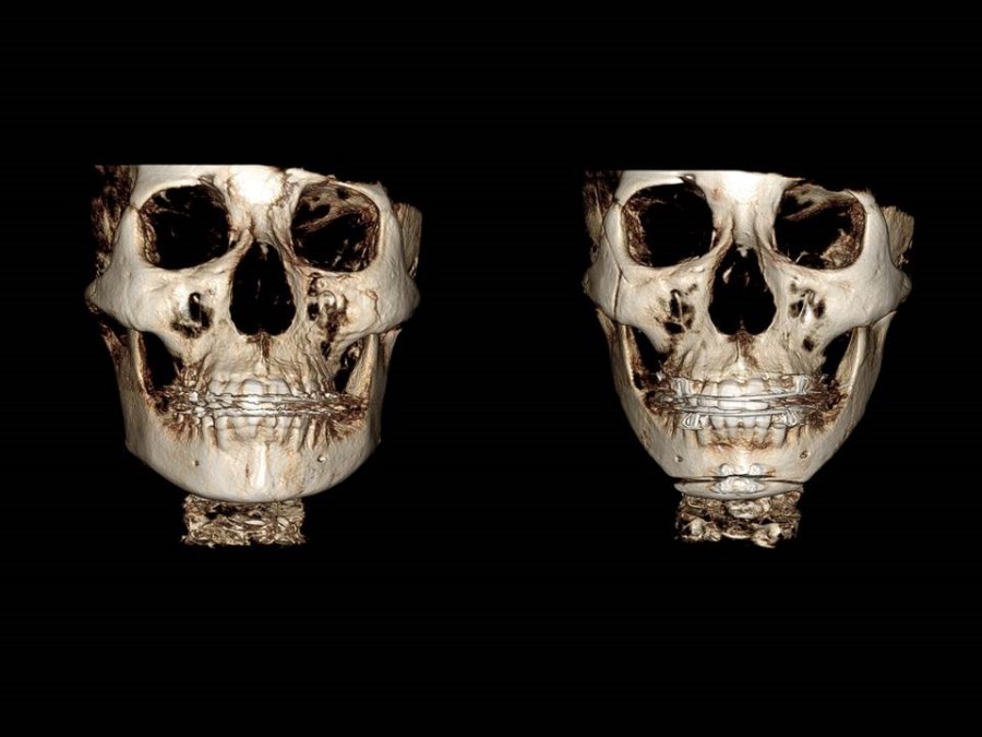
The before-and-after appearances of the cheekbones in the surgeries I performed are as follows.
The broad side cheekbone was fixed after the most posterior osteotomy of the cheekbone arch.
The continuity of the connection from the front cheekbone to the side cheekbone is preserved, and the cheekbone arch was osteotomized at the most posterior area and fixed in the correct position.
In general, the posterior area of the cheekbone arch becomes firmly united 4 to 6 weeks after surgery, and the metal fixation device can be removed if needed.
These are the before-and-after changes in the cheekbones of the person on whom I personally performed live surgery for cheekbone reduction surgery at the Korean Society of Aesthetic Plastic Surgery held at Asan Medical Center in 2012.
The position of the cheekbone arch, which was spread outward toward the back, was brought inward and fixed in the correct position.
As the width of the face decreases, you can see a change to a more three-dimensional facial shape.
The width of the right side cheekbone was broader than the left, so only the right side cheekbone was reduced while the left side was left unchanged.
This is the frontal view after inward fracture of only the right side cheekbone in the same person, and square jaw surgery and chin advancement were performed together.