Hello, this is Seoul Ob Dental Hospital.
Today, we would like to introduce the case of a man in his 30s who came in for tooth pain and was found to have a benign tumor-like lesion in the jawbone around the right lower premolar.
In particular, this surgery was completed safely and without general anesthesia, while reducing anxiety through “IV sedation” = sleep dentistry.
My gums around the right lower molars feel uncomfortable
He visited us saying that the area around the right lower molar teeth felt heavy and pressure-like.
As a result of panoramic imaging and CBCT (3D CT) examination,
a well-defined radiolucent lesion was identified within the mandible in that area.
Diagnosis results
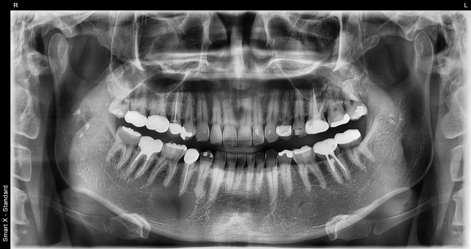
Diagnostic result: suspected benign tumor-like lesion within the mandible. The lesion was a circular cystic lesion about 2.5 cm in size, and unlike inflammation, it was a well-demarcated lesion distinguishable from the surrounding tissue.
Differential diagnosis: possibility of periapical cyst / cementoblastoma / odontogenic tumor, etc.
→ For a definitive diagnosis, lesion excision and biopsy (tissue examination) were necessary.
Treatment plan and process
Treatment method: IV sedation + lesion excision
The surgery was performed by an oral and maxillofacial surgeon, and because the patient had a fear of dental treatment,
IV sedation was selected.
✅ What is IV sedation? It is a form of anesthesia in which a sedative is administered intravenously so that the patient can receive treatment while feeling relaxed as if asleep,
while breathing is maintained.
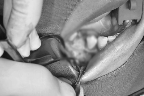
Surgical process
① Start sedation (intravenous injection) – surgery begins while the patient is comfortably relaxed
② Access to the mandibular lesion – mucosal incision and bone window creation → exposure of the lesion
③ Complete removal and curettage of the lesion – complete removal of the bony tumor tissue, including the lesion capsule
④ Guided bone regeneration and suturing – after lesion removal, allograft bone was grafted and a collagen membrane applied where needed
⑤ Pathology request (biopsy) – the removed lesion was sent to a pathologist for final diagnosis
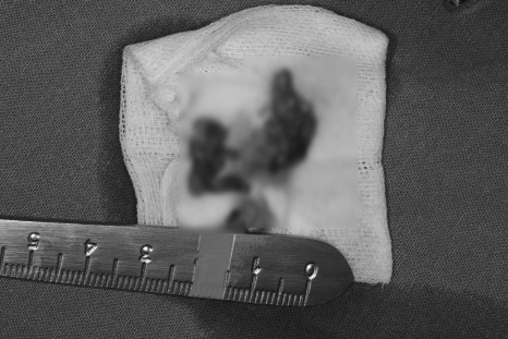
Biopsy results
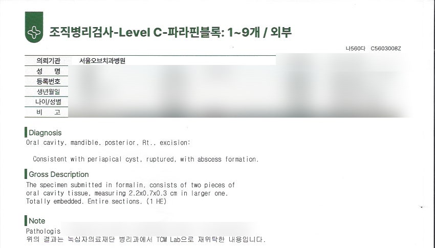
✅ Diagnosis
Oral cavity, mandible, posterior, Rt., excision:
Consistent with periapical cyst, ruptured, with abscess formation.
→ It is presumed to be a periapical cyst, and it has ruptured with abscess formation. It was confirmed that a cyst that had formed at the end of the tooth root ruptured, leading to pus formation. If a cyst becomes larger or inflammation persists for a long time,
rupture and infection may occur.
📌 Interpretation: The lesion is judged to be an inflammatory periapical cyst,
which is a representative cyst that usually forms at the root tip of a tooth due to dental infection or trauma.
Comparison before and after treatment
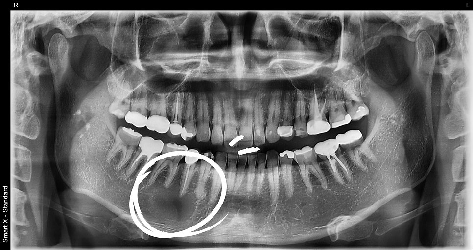
📸 Before surgery
– Cystic lesion at the periapical area
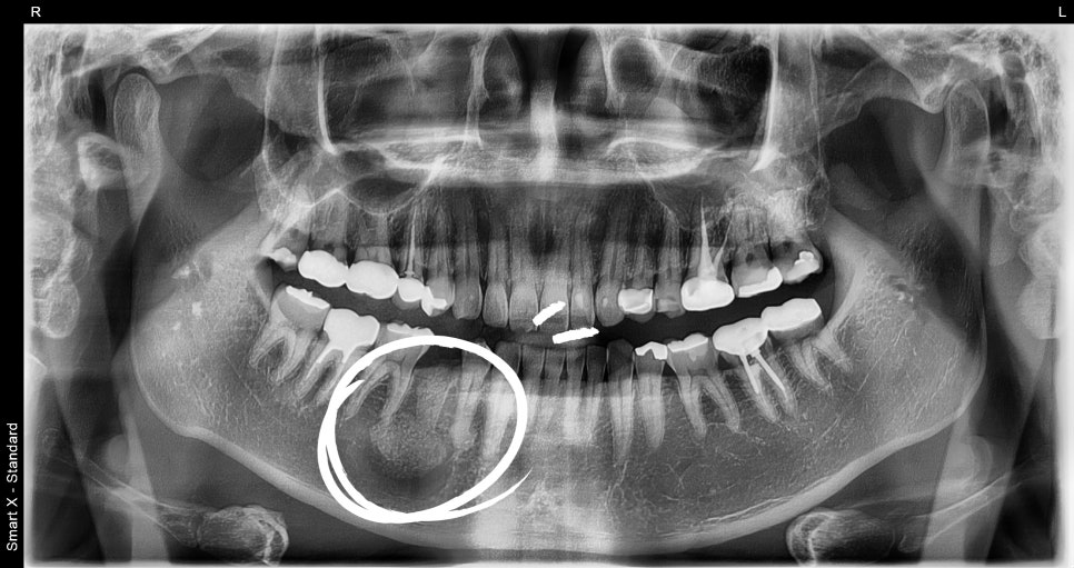
Postoperative management and results
✔️ Minimal bleeding immediately after surgery, stable recovery without complications
✔️ At suture removal after 1 week, pain and swelling were both controllable
✔️ Pathology result: Odontogenic cyst / benign tumor-like lesion
✔️ Currently being followed up with the tooth preserved
Frequently asked questions
Frequently asked questions
Q1. How do these lesions develop?
– They can mainly occur due to dental infection, trauma, or odontogenic tumors,
and although most are benign lesions, periodic checkups and early removal are necessary.
Q2. Is IV sedation the same as general anesthesia?
– Unlike general anesthesia, breathing is maintained and consciousness is partially present,
so the procedure can be received comfortably and same-day discharge is possible, with high safety.
Q3. Is it very painful after surgery?
– With IV sedation + precise anesthesia + postoperative medication,
most patients recover without significant pain.
Seoul Ob Dental Hospital is staffed by an oral surgery specialist from Seoul National University
who directly performs diagnosis and surgery, and provides various treatments such as IV sedation, sleep anesthesia, and local anesthesia,
so even patients with dental anxiety can undergo surgery comfortably.
If there are abnormal shadows on X-rays or jaw pain, as in a mandibular lesion,
it is most important not to leave it untreated and to receive an early diagnosis.
All photos used in this post are
case photos from actual treatment performed at Seoul Ob Dental Hospital,
and were published with the patient’s consent for the purpose of providing medical information.