If a patient visits a hospital because of the above symptoms, the doctor will use a detailed medical history to determine whether the reason the patient is seeking medical help is pain or instability. It is necessary to confirm the overall circumstances related to the injury, including when the symptoms the patient complains of began, how severe they are, and the mechanism of injury, and the presence of recurrence must be checked.
When a sprain occurs, the first step is to confirm whether there is a fracture with plain radiography. In patients without a fracture, after evaluating the symptoms, the degree of ligament injury and whether instability is present are confirmed through physical examination such as a stress test, and additional tests are performed if necessary. If chronic instability develops after a ligament rupture, plain radiography cannot determine whether there is an injury, and it can be diagnosed through stress radiography and magnetic resonance imaging (MRI).
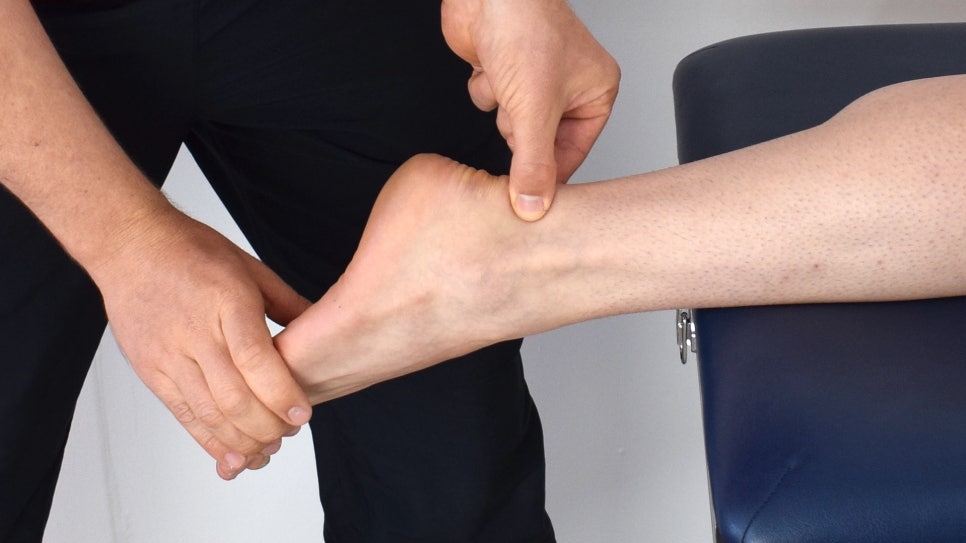
- Physical Examination
The injured area must always be examined in comparison with the normal side. Swelling, bleeding, and tenderness should be checked, and to confirm whether joint instability is present, the doctor can directly perform a stress test on the injured ligament to measure instability. In cases of sprains that occur in the ankle, knee, and thumb, a stress test of the injured ligament should be performed to measure the degree of ligament injury and the degree of joint instability.
- Radiographic Examination
Radiographic examination is widely used to diagnose bone and joint diseases and abnormalities of soft tissue. It includes plain radiography, which images the area without any special procedure, and stress radiography, which images the area while applying stress according to the injured ligament. Radiographs should be taken from the front, back, and side so that the area can be understood three-dimensionally. In growing children, because of the growth plates and bone growth depending on age, both sides must be imaged and compared.
- Plain Radiography
Ligament injuries can be diagnosed after confirming through plain radiography that there are no abnormalities in the bones or joints, such as fractures or subluxations. If a fracture occurs due to a sprain, it is not a simple sprain but a fracture.
- Stress Radiography
When a sprain occurs, plain radiography is first used to confirm that there is no fracture. This is then followed by a radiographic examination in which artificial stress is applied in the direction of the injured ligament to determine the degree of ligament rupture and the degree of joint instability.
- Magnetic Resonance Imaging (Magnetic Resonande Imaging)
Magnetic resonance imaging began to be used in the 1980s and is especially useful for diagnosis because it provides excellent contrast for soft tissue and clear anatomical differentiation. It is used very effectively not only for tumors and spinal diseases, but also for diseases affecting soft tissue such as ligaments and tendons. More recently, it has been widely used to diagnose intra-articular lesions caused by sprains by injecting contrast material into the joint cavity.
- Other Tests
Computed tomography has mainly been used to understand the exact pattern of fractures or deformities, but it has gradually come to be used for diagnostic purposes in soft tissue as well, and is especially often used for spinal diseases such as herniated intervertebral disc.
In cases of ligament injuries such as sprains, if an associated fracture is difficult to diagnose accurately with plain radiography, computed tomography is used to determine whether a fracture is present.
Ultrasound is a relatively simple test that can be performed and can diagnose ligament, tendon, and muscle ruptures without radiation exposure. However, the accuracy of interpretation varies greatly depending on the examiner, and a drawback is that accurate interpretation is difficult for anyone other than the examiner.
So far, I have explained the diagnosis of sprains.
In the next part, we will look at the treatment of sprains.
Source: Korea Disease Control and Prevention Agency National Health Information Portal