Hello,
This is Director Yeo Sang-ho of a Magok-dong dental clinic.
Many medical institutions
go through the process of
checking problems through radiographic imaging.
In this process, you may wonder:
Is radiation bad for the body?
Is it really necessary?
Because I understand these concerns well,
today I will explain the need for X-ray imaging during oral examinations
and the theoretical basis behind it.
An oral examination is
a process of discovering problems inside the mouth
that are difficult to identify by looking alone.
In this process, X-ray is also used as an important tool
to precisely evaluate the condition of internal structures such as
teeth, gums, and jawbone.
From here on, at the Magok-dong dental clinic,
I will introduce the limitations of direct observation.
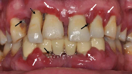
When viewed with the naked eye,
surface cavities, gum swelling, and inflammation
can be identified,
but
the tooth roots, cervical areas, and the degree of alveolar bone loss
are not easily identified.
Therefore, by using X-ray imaging,
it is possible to check abnormalities in areas not visible to the naked eye
and diagnose cavities, microcracks, nerve-related problems, and more
early.
Next, I will explain what can be checked
with X-ray at a Magok-dong dental clinic.
a. Cavities and internal abnormalities
Not only surface cavities,
but also whether decay has spread to the tooth’s pulp tissue
or to adjacent teeth
can be evaluated.
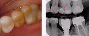
In addition,
it can be confirmed whether new decay or root inflammation
has appeared inside teeth covered by prosthetics such as
bridges or crowns.
b. Periodontal disease
When a large amount of tartar has accumulated below the gums,
which is difficult to remove with scaling alone,
its location and amount can be accurately identified
through imaging and used as consultation material.
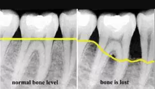
In addition,
by observing alveolar bone loss
and the inflammatory condition around the gums,
the likelihood of progression of periodontal diseases such as periodontitis
can be predicted before symptoms appear.
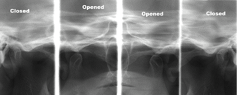
c. Condition of the jawbone and temporomandibular joint
In addition, it helps evaluate the density and height of the bone surrounding the teeth.
This is an important basis for judging the stability of natural teeth
and for establishing treatment plans such as extraction or implant procedures.
Furthermore,
structural abnormalities of the temporomandibular joint,
early arthritis, and wear can also be diagnosed,
allowing for additional physical therapy or management measures
to be prepared.
What is the principle of X-ray imaging?
At the Magok-dong dental clinic,
I will explain its characteristics and how it works.
X-ray uses high-energy electromagnetic waves
to pass through various tissues of the human body.
Depending on the density of each tissue,
the degree to which the electromagnetic waves are attenuated differs,
and teeth and bones, which have high density,
absorb a large amount of radiation,
whereas soft tissues absorb relatively little.
Thanks to this property,
teeth and bones appear bright on images,
while cavities, inflammation, and areas affected by decay appear dark.
Based on the amount of radiation
that reaches the film or digital sensor,
the bright and dark areas of the imaging region
are clearly contrasted to create an image,
and through this,
specialists can analyze the density of each tissue
and any structural abnormalities.
How is it reflected in diagnosis?
At the Magok-dong dental clinic and all oral medical institutions,
treatment plans based on X-rays
can be established as follows.
✔ Preventive measures
By continuously monitoring tartar below the gums and changes in bone,
preventive measures are implemented before problems worsen.
✔ Decision on additional treatment
By comprehensively evaluating the condition of prosthetics and the overall condition inside the mouth,
additional care such as prosthetic replacement or implant procedures
is decided when necessary.
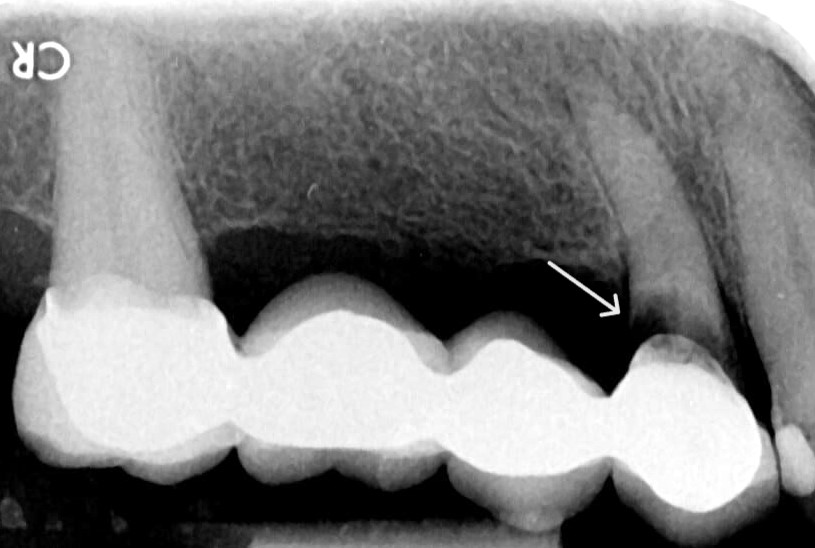
✔ Confirmation of treatment history
Past treatment records can also be understood by sight,
but for structures that cover the entire tooth, such as crowns,
it may be necessary to confirm whether there are internal abnormalities.
Today, I explained the need for X-ray imaging during oral examinations
and its theoretical basis.
I hope this was helpful to those who needed medical information related to this topic.
This was Director Yeo Sang-ho of a Magok-dong dental clinic.
Thank you for reading this long post.