Hello. I am Director Kim Jun-hyeon.
A patient came to the clinic with concerns about both a deviated septum on the inside and the appearance of the nose.
I would like to introduce a case in which we achieved both cosmetic improvement of the nose and functional improvement as well.
Crooked nose correction
✓ Crooked nose
✓ Hump nose
✓ Low nasal bridge
✓ Drooping nasal tip
✓ Functional nasal obstruction (deviated septum)
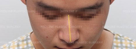
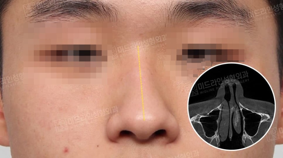
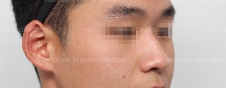
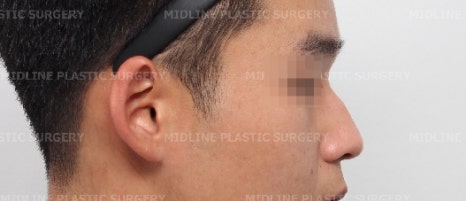
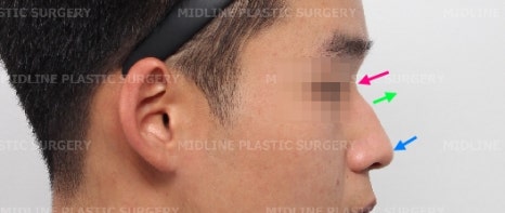
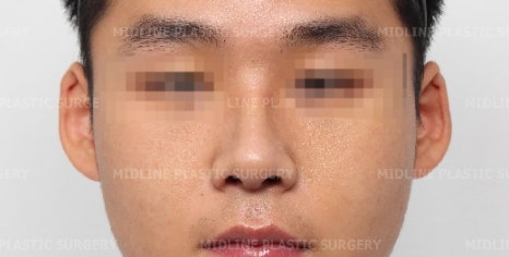
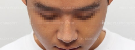
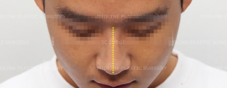
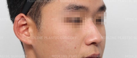
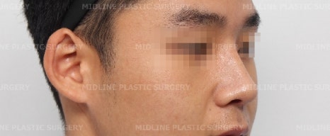
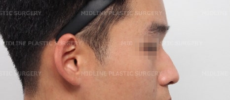
Let me explain while looking at the photos before surgery.
First, let’s look at the cosmetic aspects.
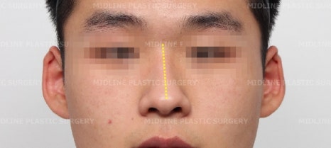
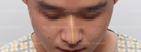
✓ Crooked nose
When viewed from the front and when the head is tilted downward, the direction of the curve is marked in yellow.
✓ Low nasal bridge ✓ Drooping nasal tip
The nasal bridge between the eyebrows was also low, so it needed to be raised.
The low bridge is marked in fluorescent purple.
Also, the drooping nasal tip that needed to be lifted is marked with a blue arrow.
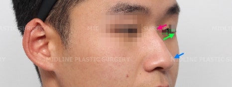
✓ Hump nose
The hump that needed to be removed is marked with a fluorescent light-green arrow.
Q. If the nasal bone is crooked, is the septum also crooked?
Some people ask this.
A. A crooked nose means the external bone structure is crooked, and even people whose noses do not appear crooked can have a deviated septum.
That is why, at our clinic, for a detailed diagnosis, we perform CT scans not only for crooked noses, but also during overall nose consultations for low noses, bulbous noses, and more.
This helps us assess the functional aspect based on the degree and direction of septal cartilage deviation, while also checking the size of the septal cartilage to be used for the nasal tip and confirming the structure of the hump.
The septum is located at the center, dividing the inside of the nose into left and right sides, and its front part is cartilage while the back part is bone.
By performing a CT scan from the front part to the back part, we can determine the cause of nasal obstruction and any functional abnormalities.
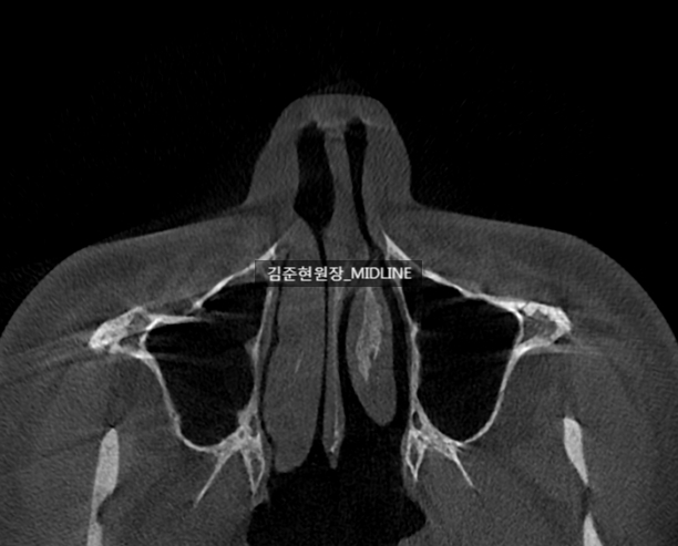
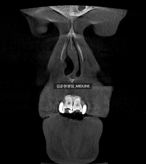
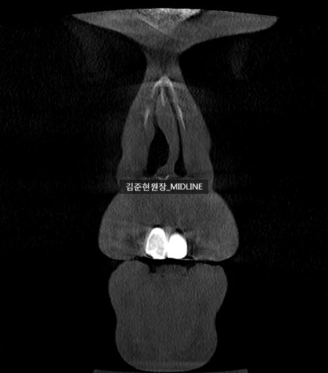
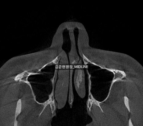
CT with a deviated septum (axial view)
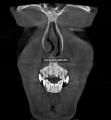
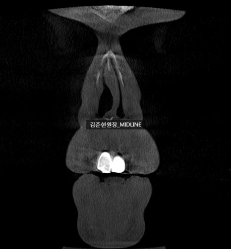
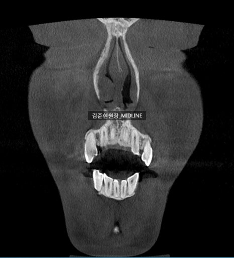
CT with a deviated septum (coronal view)
In the CT above, the cartilage was larger than the septal space, and the lower part of the cartilage was bent to the left, showing a deviated septum.
If the nose is raised while leaving such a septum untreated, there is a very high chance that the nose will collapse to one side or the columella will become crooked.
So, using the Swing door technique described in a previous post,
we performed caudal septal relocation.
https://blog.naver.com/creating_beauty/222181570405
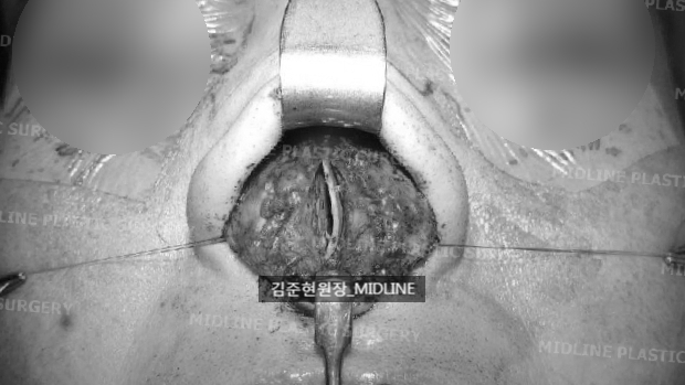
Here is a photo of the severely deviated septal cartilage removed during surgery.
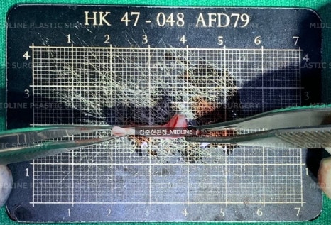
The removed septal cartilage shows the same curvature seen on the CT.
This is the posterior cartilage of the septum, and to prevent the nose from collapsing (and to prevent saddle nose deformity),
we need to preserve 8–10 mm of the anterior portion of the entire septal cartilage.
In other words, only the portion marked with the fluorescent purple arrow in the figure below should remain.
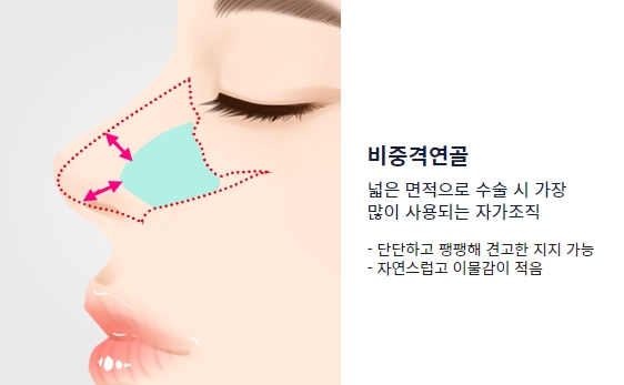
Excerpt from the Midline Plastic Surgery website
https://midlineps.com/nose/
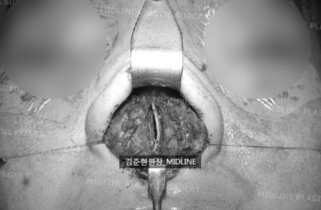
This is the septum after leaving a safe amount in place. Before caudal septal relocation, it is still crooked.
Crooked septal cartilage
The lower part of the septal cartilage is separated, and the cartilage extending beyond the space where the septum should be is removed.
Then it was re-fixed to the anterior nasal spine, the bone at the very front where the septal cartilage is attached.
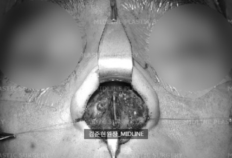
The septal cartilage straightened after caudal septal relocation using the Swing door technique
Through caudal septal relocation,
we corrected the crooked anterior septum,
and even when a septal extension graft was later placed using the harvested septum to lengthen and heighten the nose,
we created a stable foundation that would not bend.
In this way, while performing functional rhinoplasty,
we also completed cosmetic rhinoplasty, including hump correction, crooked nose correction, and bridge augmentation.
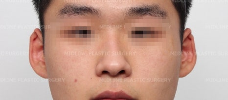
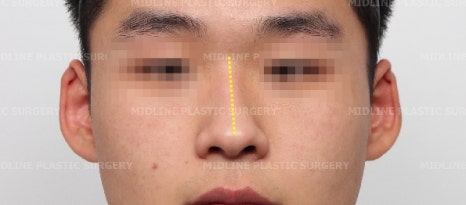
Here are the before-and-after photos.
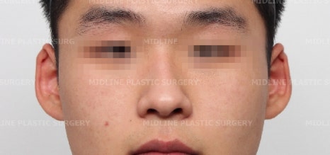
If you look at the front view, you can see that the crooked nasal bridge was corrected, improving the crooked nose.
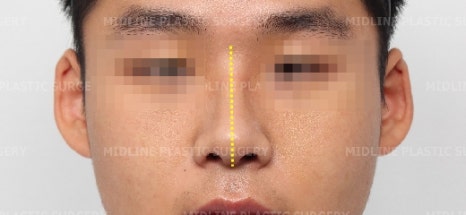
As before, I marked the direction of the nasal deviation in yellow.
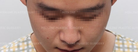
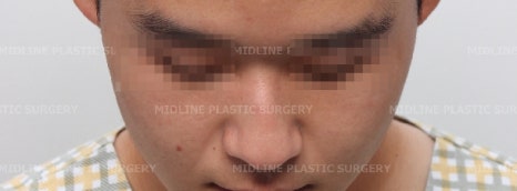
You can also see the corrected result when the head is tilted downward.
You can also see the improvement in the low nasal bridge, the hump correction, and the drooping nasal tip.
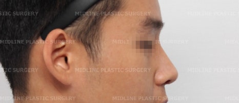
The surgery was successfully completed this way.
We also confirmed through CT how the functional issue involving the deviated septum had been corrected.
For easier comparison, I captured images from the same positions.
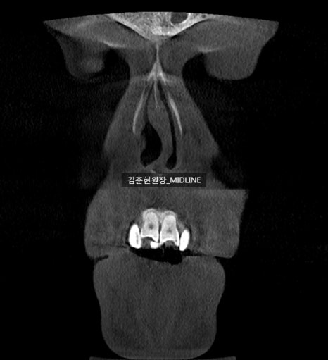
CT before surgery showing a deviated septum (coronal view)
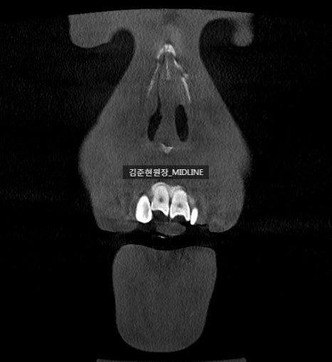
Corrected septal CT after surgery (coronal view)
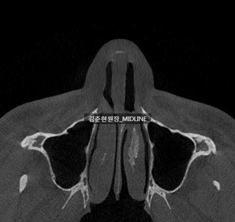
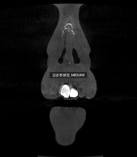
Left) Preoperative CT with deviated septum / Right) Postoperative corrected septal CT (axial view)
Left) Preoperative CT with deviated septum / Right) Postoperative corrected septal CT (axial view)
Today, I showed you a case in which functional nose surgery and cosmetic nose surgery were performed together.
-
The septal deviation extended to the front part of the cartilage, so it was corrected.
-
There was a hump, so it was corrected.
-
The crooked nose was straightened through osteotomy.
This was the photo taken one week after surgery, and there was almost no bruising or swelling, so the patient was also surprised. ^^
<If you have any additional questions, please leave a private comment and I will kindly answer them. Thank you.>
.
.
.
.
.
.
.
.
.
.
I also attached videos taken from different angles before and after surgery.