Dark circles under the eyes can be caused by congenital factors or can develop as part of the aging process.
People usually begin to notice changes under the eyes in their 30s and 40s, when aging around the eye area starts, so interest in lower eyelid fat repositioning surgery is high.
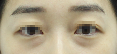
Dark circles under the eyes in a person in their late 30s
When dark circles appear under the eyes, it is easy to start considering various procedures.
In general, it may be possible to address them relatively simply by injecting filler or collagen injections into the hollowed area, but the maintenance period is short, and there are many cases where the result is not satisfactory.
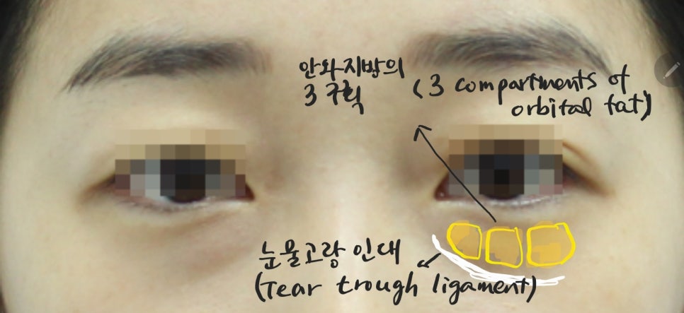
The compartments of lower eyelid fat and the tear trough ligament
The lower eyelid fat is divided into three fat compartments, called the medial, central, and lateral compartments.
If the fat pad surrounding this orbital fat becomes loose, the lower eyelid fat may protrude, and a boundary is formed with the tear trough ligament under the eye, making the under-eye area look older.
Because lower eyelid fat repositioning surgery is performed through the conjunctiva, it has the advantage of no external incision, but the surgical difficulty is not low because the field of view is narrow.
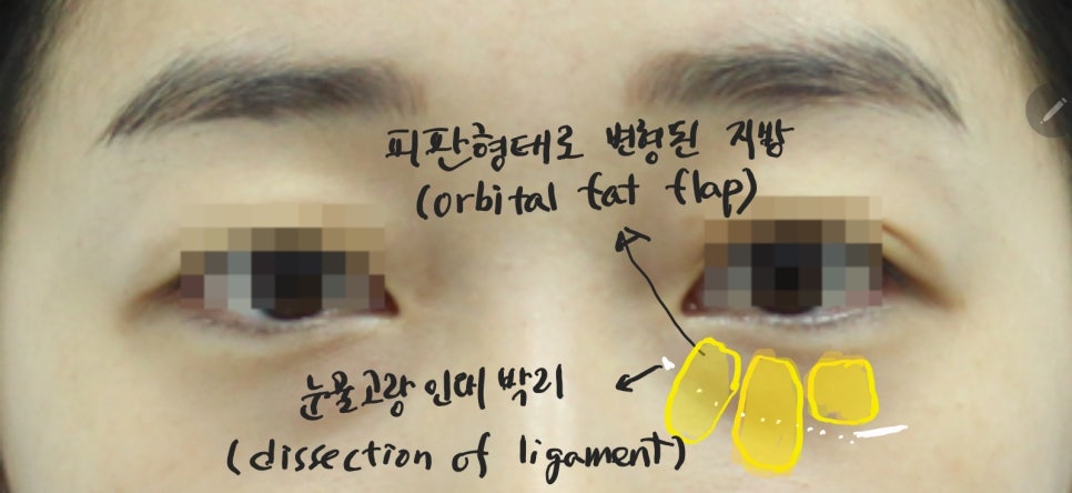
Dissection of the tear trough ligament and changes in the flap shape of the lower eyelid fat
Removing only the lower eyelid fat is one possible method, but that approach often leads to frequent recurrence or disappointing results that make the under-eye area look hollow.
First, the tear trough ligament that creates the under-eye tear trough must be dissected. Since this is a firm tissue, it must be carefully dissected at the exact location, and it should be dissected sufficiently downward under the eye for the best effect.
Then the lower eyelid fat is carefully separated and made into a long flap. This is necessary to improve the survival rate of the repositioned fat and to secure continuity with the original lower eyelid fat, allowing for a natural change in the under-eye area.
(Flap: when tissue is moved, it is transferred in an extended form so that blood circulation can be maintained)
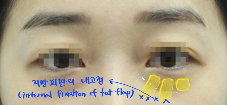
Internal fixation of the lower eyelid fat flap
The dissected fat flap is moved downward and fixed in place. Because this process is performed through a narrow field of view, sufficient visibility must be secured without bleeding.
The method used to fix the flap should also be internal fixation. With external fixation, once the sutures are removed within a week, the flap may return to its original position.
When performing internal fixation of the fat flap, it should be secured to the lower eyelid periosteal area with a strong absorbable suture so that the fixation lasts for at least about one month, allowing the moved fat flap to remain in place instead of returning to its original position.
(Internal fixation method: a method of fixing the fat flap internally to the periosteal area using absorbable sutures)
(External fixation method: a method of exposing the sutures outside the skin with non-absorbable sutures and removing them after one week)
If you look at the surgical diagram for lower eyelid fat repositioning from a side cross-sectional view, it is as follows.
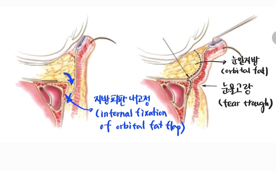
Side surgical diagram of lower eyelid fat repositioning
It shows the hollow tear trough being sufficiently dissected, and the bulging fat pad above being lowered in the form of a flap and internally fixed from within.
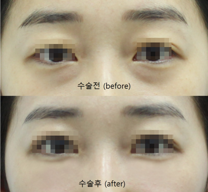
Before-and-after comparison
Before surgery, the bulging fat under the eyes and the tear trough are neatly arranged without any uneven contour.
Lower eyelid fat repositioning is a procedure with the advantage that, when performed well, the results are good and long-lasting.
The removal of lower eyelid fat is the least desirable method, and even if the surgery is complicated, it is best to reposition the fat using an internal fixation method in flap form.
In addition, because the surgery is performed through the conjunctiva, there is no external incision, no need for suture removal, and recovery is quick.