After rhinoplasty, there is a tendency for the tip of the nose to drop again due to the pressure of the stretched skin.
Therefore, to secure sufficient support for the nasal tip, a strong support structure should be built using septal cartilage, ear cartilage, and similar materials.
Some people visit the hospital for revision surgery because of a lowered nasal tip, but sometimes we see patients whose nasal tip has dropped severely.
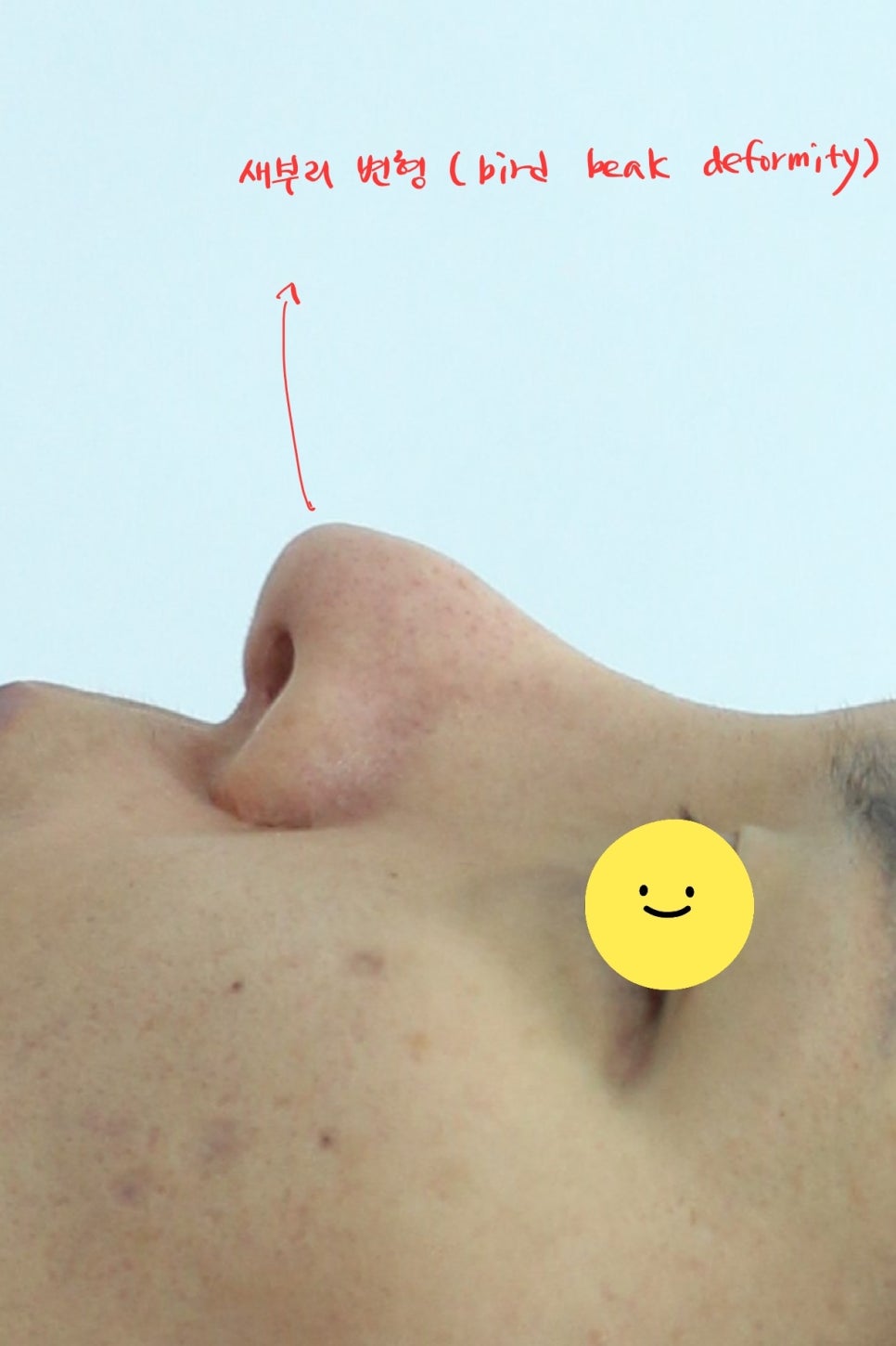
Bird beak deformity with a lowered nasal tip height
The patient said that after undergoing rhinoplasty with donated rib cartilage in the past, the nasal tip had become significantly lower over time.
Donated rib cartilage can be a good material when used appropriately, but it is always necessary to keep in mind that there is some possibility of resorption.
Also, because donated rib cartilage is not autologous cartilage, it may be resorbed, and in some cases it can break due to calcified areas.
For that reason, rather than building the nasal support structure using only donated rib cartilage, it is better to use it as an auxiliary material along with septal or ear cartilage.
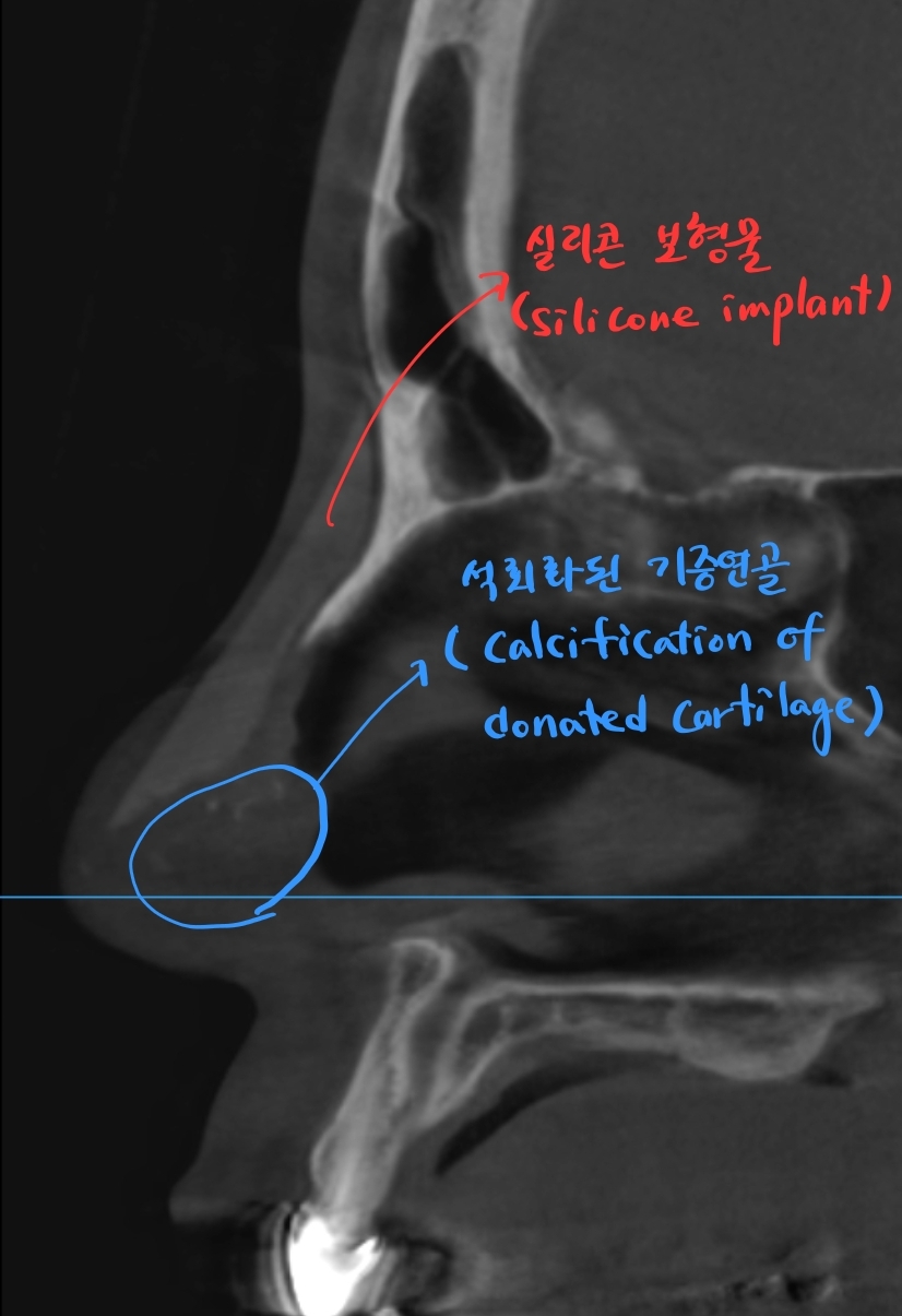
Calcified areas of donated rib cartilage visible on preoperative CT scan
The bridge contains a silicone implant of about 6 mm in height, and slightly faint calcified areas are visible at the nasal tip, which are presumed to be calcified portions of donated rib cartilage.
In the end, although the nasal tip had been raised with donated rib cartilage, it is highly likely that the cartilage was resorbed and the calcified portions weakened, resulting in insufficient support for the nasal tip.
As a result, the nasal tip collapsed and came to look like a bird beak.
The height and shape of the bridge implant also seem somewhat less than ideal.
First, the silicone implant and donated rib cartilage that were in the nose are removed.
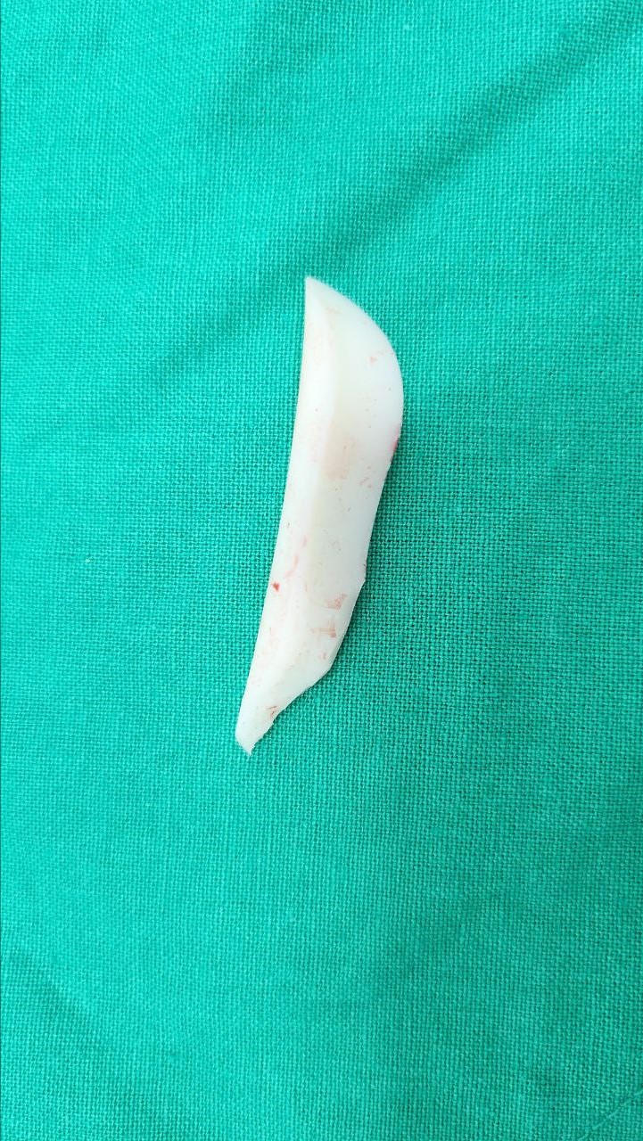
Removed silicone implant
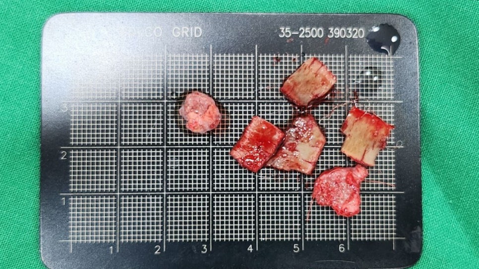
Removed pieces of donated rib cartilage
Donated rib cartilage was used as support for the nasal tip, but because of the calcified areas, it often crumbles during removal.
Ultimately, to secure sufficient support for the nasal tip, autologous rib cartilage was harvested for the rhinoplasty.
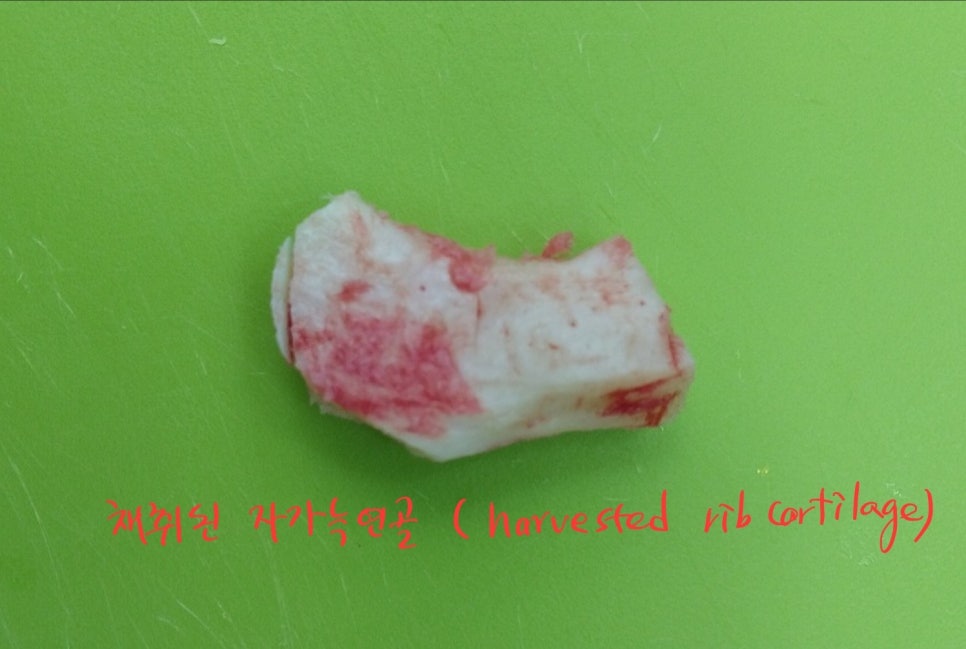
Harvested autologous rib cartilage
When using autologous rib cartilage for the nasal tip, it must be processed into pieces because it is used by extending the existing septum or as a support structure.
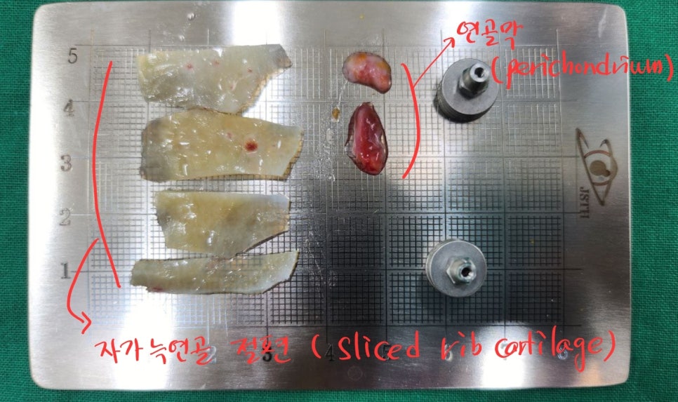
Rib cartilage pieces and perichondrium
The reason rib cartilage is made into multiple pieces is because of its tendency to warp. Since rib cartilage tends to bend to one side, it must be used by balancing the forces and attaching pieces on both sides so that the nasal tip does not bend after surgery.
In addition, the perichondrium around the cartilage is also valuable material. In revision rhinoplasty, areas of the skin may become thin for various reasons, so it may be necessary to graft perichondrium to appropriate areas.
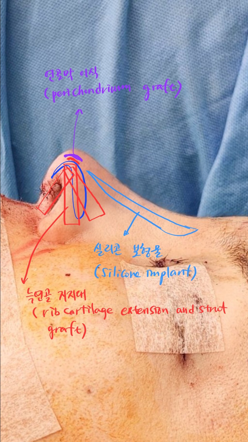
Diagram of autologous rib cartilage revision rhinoplasty
It is also important to graft the autologous rib cartilage nasal tip support structure in both directions to prevent warping, and it must support the nasal tip from multiple directions for sufficient support to be achieved.
Before surgery, the nasal tip had collapsed and the nose had changed into a bird-beak shape, but after surgery, the nasal tip support was secured and the line became more feminine.
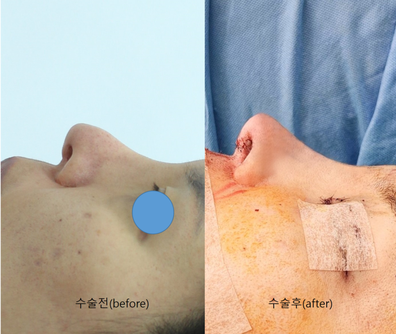
Before-and-after comparison
Donated rib cartilage is a material used when there is not enough autologous cartilage, but because of the nature of the product, there may be products with a high resorption rate and a high proportion of calcification, so this must always be kept in mind during surgery.
Therefore, rather than building the nasal tip support structure using only donated rib cartilage, it is better to use it as an auxiliary material along with septal cartilage or ear cartilage, which increases predictability after surgery.
Also, the safest cartilage with the best support and the highest predictability after surgery is autologous rib cartilage.
If you are considering revision surgery because of the problem of the nasal tip dropping after rhinoplasty, it would be a good idea to look into autologous rib cartilage rhinoplasty.

Bird-beak nose review. This is a real model who agreed to disclosure.