For those who visit for a consultation for revision rhinoplasty, information about prior surgery can be gathered through a medical interview, but many people do not remember their past surgery accurately.
Because of this, CT scanning is often performed during the consultation process.
Although a CT scan may be an inconvenient process, it provides a lot of useful information, so we recommend it for preoperative diagnosis.
The most basic information that can be obtained through a CT scan is the shape and height of the implant inserted into the bridge of the nose in a previous nose surgery.
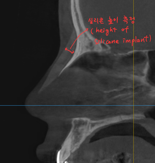
Shape and height measurement of the existing implant
By evaluating the height of the existing implant, a surgical plan can be made regarding whether to raise the nasal bridge higher than it is now, create a similar height, or lower it slightly.
If the height of the nasal bridge and the shape of the implant are appropriate, it may also be possible to evaluate whether revision surgery can be limited to the nasal tip.
Usually, as in the case above, the implant is mostly an I-shaped silicone implant, but in some cases an L-shaped silicone implant is used.
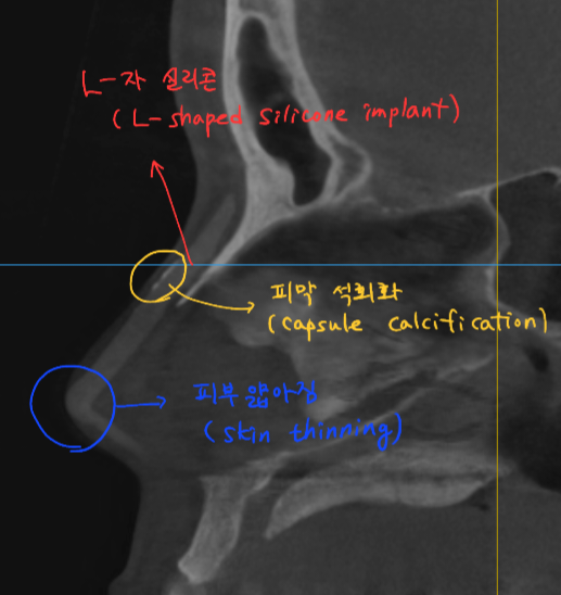
L-shaped silicone implant
In the case of L-shaped silicone, it usually does not sit closely against the nasal bridge, so there are often many empty spaces and it can shift.
Also, if a long time has passed after surgery, a thick capsule around the silicone implant or calcification of the capsule may sometimes be found.
However, the biggest drawback of L-shaped silicone is that because the silicone implant extends all the way to the nasal tip, it thins the skin at the tip.
Therefore, if L-shaped silicone has been inserted, additional consultation may be needed regarding methods to reinforce the skin at the nasal tip.
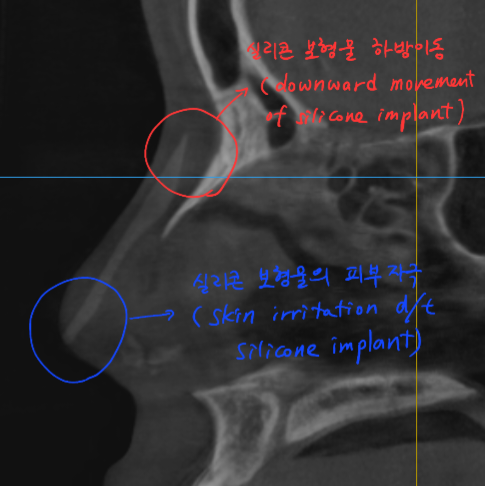
Downward displacement of the silicone implant
In some cases, even when rhinoplasty was performed using an I-shaped silicone implant, if there is chronic swelling and inflammation or if contracture gradually develops, the silicone implant may move downward toward the nasal tip and irritate the skin there.
Another piece of information that can be obtained through CT scanning is the internal nasal environment, which may be hidden by the silicone implant.
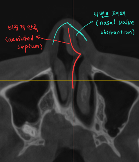
Deviated septum and internal nasal valve narrowing
After rhinoplasty, we sometimes see cases where the nose appears crooked, the nostrils become asymmetrical, or nasal congestion develops.
In such cases, a CT scan may show that the deviated septum has worsened or that one internal nasal valve is blocked, causing nasal obstruction.
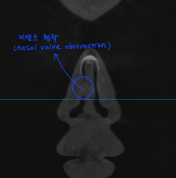
Internal nasal valve narrowing
Because the nose is located at the center of the face, it is important not only that it does not look crooked from the outside, but also that after rhinoplasty it does not cause discomfort by having a deviated septum or causing nasal congestion.
Finally, another important piece of information obtained through CT scanning is assessing the size of the remaining septal cartilage or identifying the materials previously used in the nasal tip.
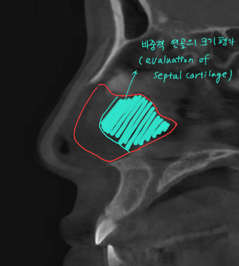
Assessment of the amount of septal cartilage that can be harvested
If rhinoplasty in the past was performed without using septal cartilage, the size of the septal cartilage can be estimated to some extent through CT scanning. This can help predict how much the nose can be raised or help plan whether additional cartilage harvesting will be needed.
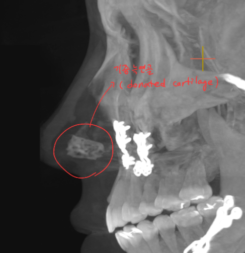
Donor rib cartilage used in the nasal tip
It also allows us to know what material was used in the nasal tip during prior surgery, so we can plan which cartilage to use as material for the next rhinoplasty.
The information that can be obtained through CT scanning during a consultation for revision rhinoplasty is very diverse, beyond the items listed above.
However, some people sometimes worry that getting a CT scan exposes them to unnecessary radiation.
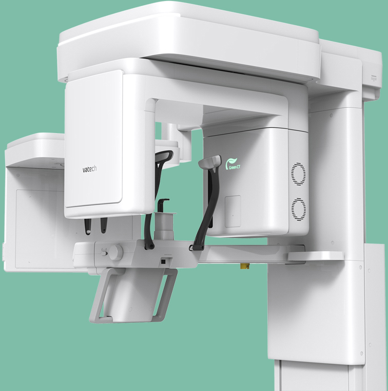
Low-dose Green CT
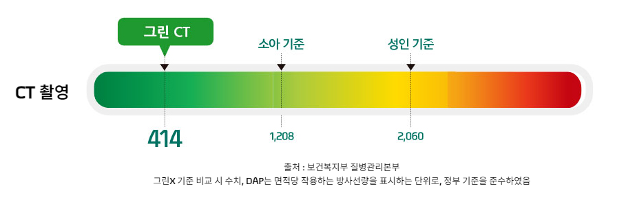
At our hospital, we have installed a latest-generation low-dose Green CT that significantly reduces radiation exposure compared to conventional CT, helping to ease concerns about radiation exposure.
When addressing previous concerns during a revision rhinoplasty consultation, it is important to establish a good surgical plan, and CT scanning can provide a great deal of information about prior nose surgery and the condition inside the nose.