
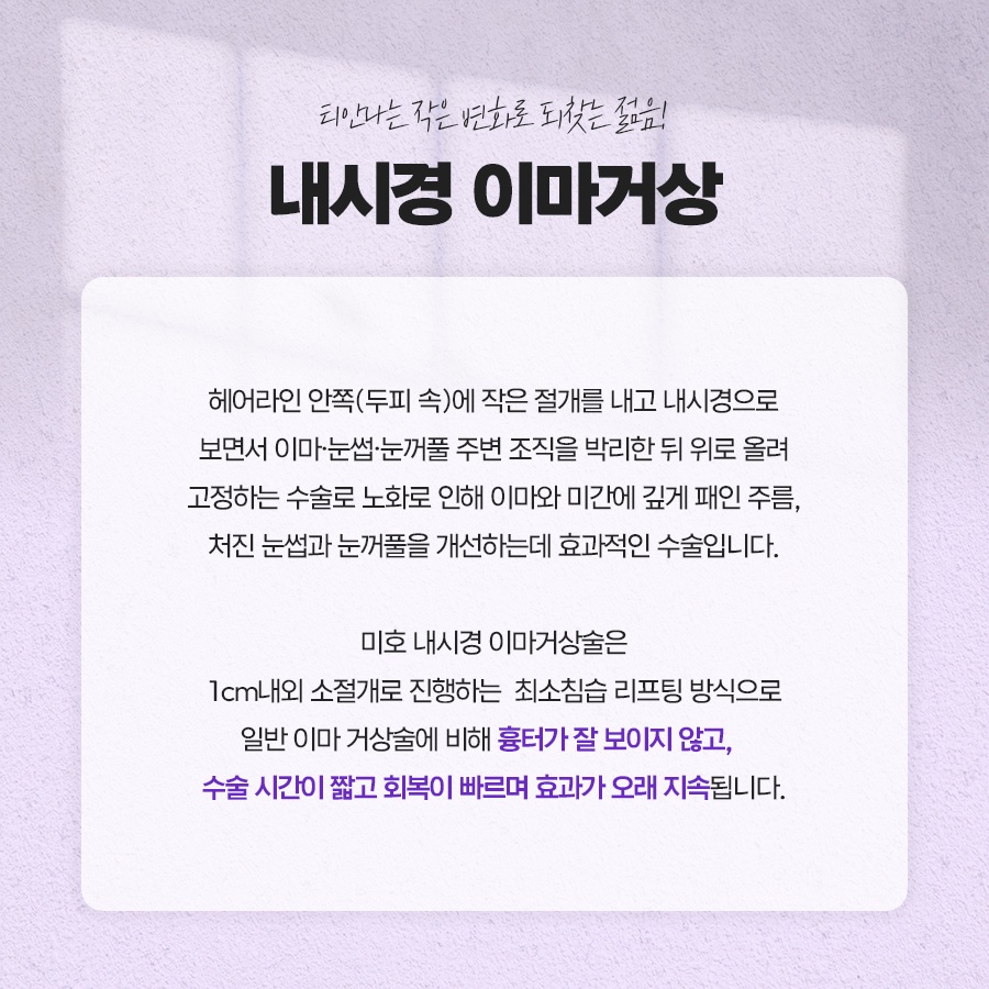
As people age, many begin to feel that their eyelids have become heavy and that their forehead wrinkles have deepened.
Because the upper face greatly influences one’s overall impression, even small changes can make a person look older.
Noninvasive methods such as fillers or Botox have limitations when it comes to restoring tissue that has already descended, so many people seek a more fundamental approach.
Against this backdrop, interest in endoscopic forehead lifting continues to grow.
Today, we will look at the principles by which this method improves sagging areas and examine the recovery process.
The forehead and the area around the eyebrows gradually descend over time.
As the soft supporting structures, including the skin and ligaments, weaken, the effects of gravity and downward muscle activity add to the changes, making the eyelids feel increasingly heavy.
As these changes accumulate, the face may look tired or constricted even at rest, and some people develop a habit of using their forehead muscles when opening their eyes.
The principle of endoscopic forehead lifting focuses on returning these downward-shifted structures to a higher position.
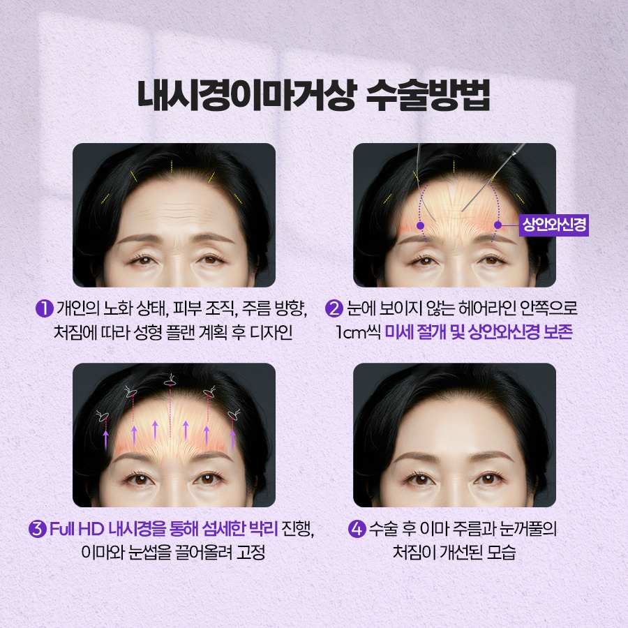
The basic process involves making several small openings in the scalp, inserting a thin camera, directly examining the area beneath the skin, and performing the correction.
Several access channels measuring approximately 1.5–2 cm are created behind the hairline. The skin and muscle layers are carefully separated beneath the periosteum, after which the entire loosened tissue is lifted upward and secured.
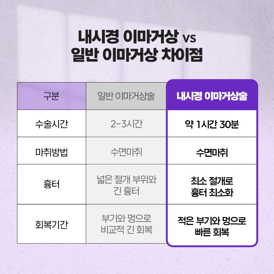
The endoscopic forehead lift differs from conventional incision-based methods in that it can address a wide area without a large incision. This helps reduce the concern many people have about scarring while maintaining the lifting range.
The following are examples of cases in which this method may be considered.
Typical examples include deeply etched forehead wrinkles and brows that have descended over the eyelids, making the field of vision feel restricted.
It may also be an alternative for people who have developed a habit of frequently frowning between the eyebrows, giving them a stern appearance; those who have repeatedly received Botox but feel that its duration of effect has gradually shortened; and those whose naturally low eyebrow position often makes them look angry when their face is expressionless.
Because suitability varies depending on the individual’s condition, a preliminary consultation is important.
The location of the incisions is one of the factors that affects the naturalness of the result.
Three to five short openings are made inside the hairline and hidden within the scalp, making them difficult to see from the outside.
Each incision is generally no longer than 2.5 cm. When made in a zigzag pattern, follicle damage is reduced, allowing the hair to grow back naturally during recovery.
Compared with older methods that involved making a large incision along the frontal hairline, this approach generally places significantly less burden on scarring.
In addition, the incision sites are positioned away from the paths of the nerves responsible for scalp sensation, helping to reduce recovery-period discomfort such as decreased sensation.

After the sagging areas are lifted upward, the tissues are secured in place to maintain their position stably.
Endotine is commonly used. It is an absorbable material with pyramid-shaped projections that supports the tissue by gripping it, and it gradually dissolves in the body within six months to one year.
A bone-tunneling technique may also be used, in which small tunnels are created in the bone and sutures are passed through them.
The fixation methods used in endoscopic forehead lifting can be combined in various ways and differ according to each individual’s condition.
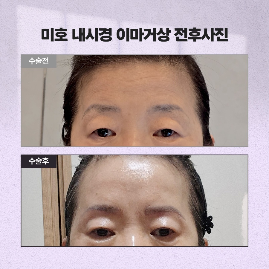
Muscle adjustment is another area addressed along with the correction of sagging.
The corrugator muscles, which create vertical lines between the eyebrows, and the procerus muscles, which create horizontal lines above the bridge of the nose, repeatedly contract with facial expressions and contribute to the formation of deep creases.
The depressor supercilii muscles, which pull the eyebrows downward, and the upper part of the orbicularis oculi muscles may also be addressed. The balance among these muscles must be maintained for the eye contours to settle at a natural height.
The goal is to reduce the strength of each area only as much as necessary, thereby reducing how often new wrinkles form.
However, excessive adjustment can make facial expressions appear unnatural, so the medical professional’s judgment regarding how far to adjust the muscles has a significant impact on the result.
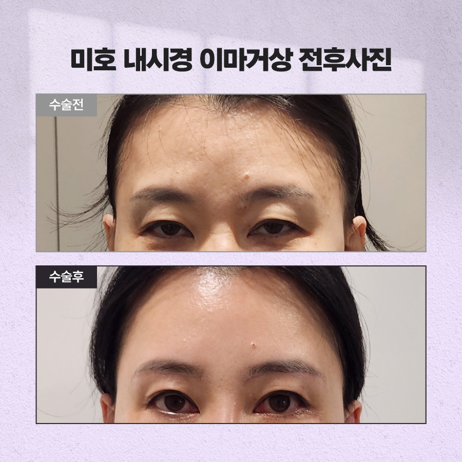
Next, let us look at the recovery process step by step.
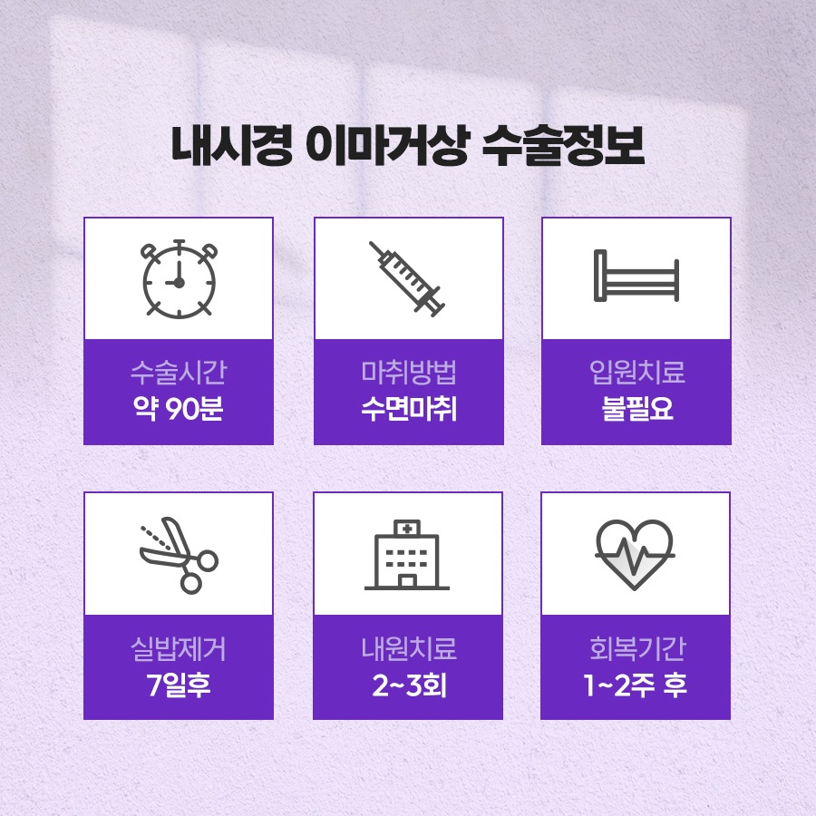
Swelling is generally most pronounced around 72 hours after the procedure and then visibly subsides over the following one to two weeks.
The swelling may spread downward and temporarily cause bruising around the eyes, while sutures are usually removed after about one week.
The scalp may feel numb or tingly for some time, but as the nerves regenerate, these sensations generally begin to improve gradually after two to three months.
Individual differences should be taken into account during recovery from an endoscopic forehead lift.
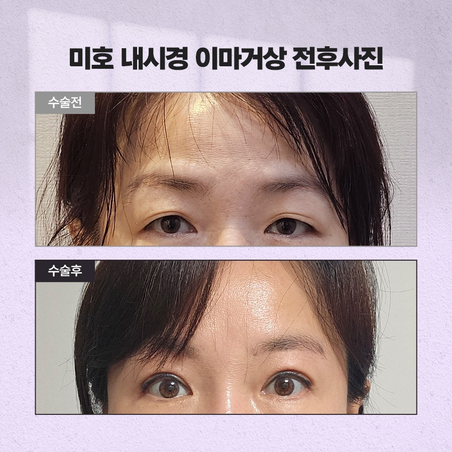
Next, we will explain appropriate care methods in detail.
During the first three days or so, frequent cold compresses may help relieve swelling and pain. It is also advisable to avoid facial expressions that involve extensive movement of the forehead muscles for the time being.
Hair washing is generally possible after two to three days using a mild shampoo. Since drinking alcohol and smoking can delay recovery, it is safer to avoid them for approximately two to four weeks.
Saunas and strenuous exercise are best resumed gradually after three to four weeks. If fever or persistent pain occurs, notify the medical professional immediately.
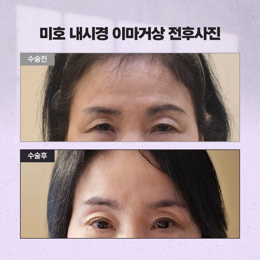
Endoscopic forehead lifting is not completely free from the possibility of complications.
In rare cases, temporary reduction in eyebrow movement may occur, and decreased sensation or asymmetry between the two sides may also develop.
Temporary hair loss around the incision sites may occur, and additional treatment may be necessary due to bleeding-control issues. Depending on skin elasticity and the strength of fixation, the tissues may also become loose again.
Therefore, a safe decision involves discussing your degree of sagging and expectations sufficiently and consulting with a medical professional who has an in-depth understanding of anatomy.



Previous image
Next image