Hello!
I am the director of Seoul Bardi Dental Clinic.
▼ Check the Dental Clinic Location ▼
| This post was written directly by Seoul Bardi Dental Clinic in compliance with Article 56, Paragraph 1 of the Medical Service Act for the purpose of providing medical information. The information provided is for reference only. We recommend visiting a medical institution and receiving guidance from a medical professional for symptom assessment and accurate management methods. All procedures and surgeries performed at a dental clinic may involve risks depending on the individual, such as inflammation, bleeding, and swelling. Please make your decision after sufficient consultation with a medical professional. All images were created using AI to aid understanding. |
Many people brush their teeth well
and receive regular checkups,
yet one day they visit because a tooth has suddenly broken.
In most cases, the cause is the same.
Not a cavity on the chewing surface,
but a root surface cavity that developed at the boundary between the gums and teeth.
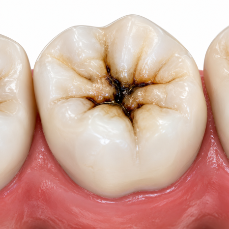
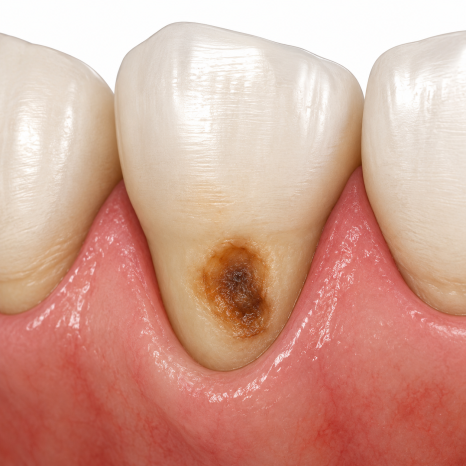
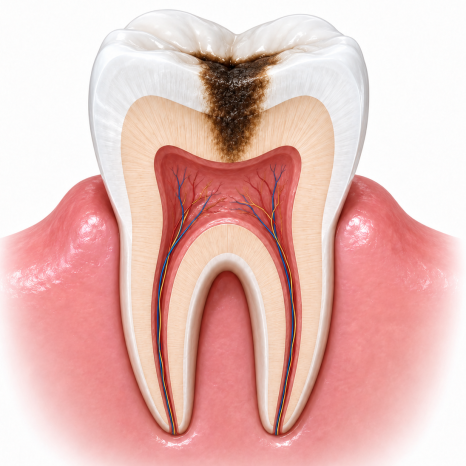
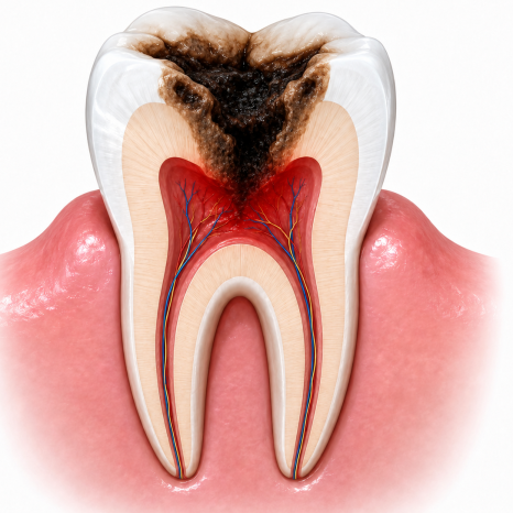
Example photos
About one-third of people aged 65 and older have them,
and they are common cavities found in approximately 31.9% of adults as well,
yet most people do not realize they have them.
(Zhao Y et al., 2023, BMC Oral Health)
Today, I will explain why root surface cavities are more dangerous than ordinary cavities,
what signs can help you notice them,
and how to prevent them.
| Directions to the Clinic |
| Root Surface Cavities: Why Do They Progress More Quickly? |
Even when the cavity is the same,
its rate of progression can vary completely depending on its location.
The outside of a tooth
is covered with enamel, the hardest outer layer,
while the inside is dentin, the softer layer surrounding the nerve.
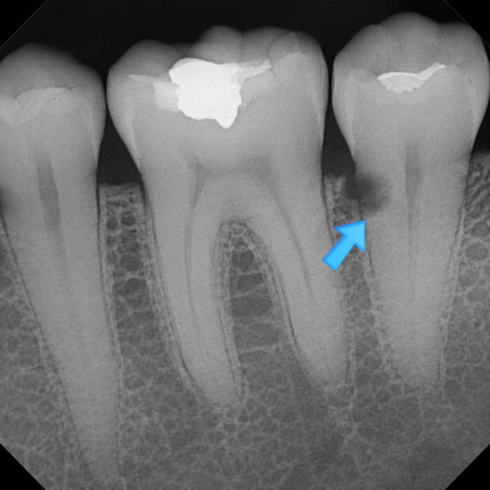
Example photo
A cavity on the chewing surface must penetrate the enamel
before it can reach the nerve.
In contrast, a root surface cavity begins in the dentin.
This is because the tooth roots beneath the gums do not have enamel.
Dentin is weaker than enamel.
Its mineral content is approximately 70%—lower than enamel's approximately 96%—
and its pH threshold for beginning to dissolve in acid is also higher.
Simply put, this means it breaks down more quickly even when exposed to a weaker acid.
Even when exposed to the same amount of acid,
a root surface cavity reaches the nerve more quickly.
(Frontiers in Oral Health, 2025 / Zhang et al., 2020)
| Why Are They Difficult to Notice? |
Three reasons overlap.
- The location cannot be seen
They progress inside the gum line and cannot be seen in a mirror.
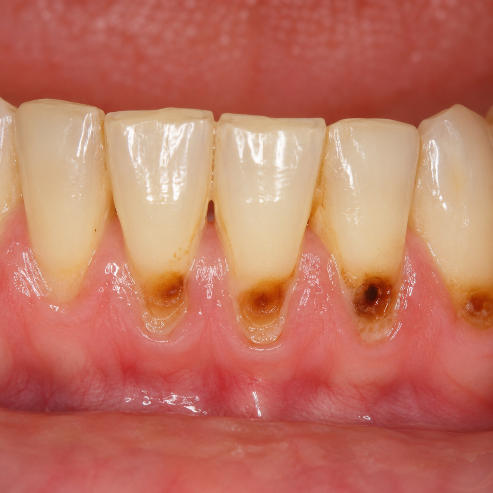
Example photo
- Pain comes late
Dentin contains tiny tubes called dentinal tubules,
so at first there may only be slight sensitivity,
with pain beginning when the cavity has nearly reached the nerve.
- They occur along with gum recession
As people age, the gums naturally recede,
and the exposed root surface is immediately placed at risk of developing cavities.
The gums may also recede slightly and temporarily after scaling.
(Hayes M et al., 2016, J Dent — Analysis of root surface cavity risk indicators)
People aged 65 and older who are more likely to develop root surface cavities
If any of the following apply to you, it is advisable to have regular checkups
at somewhat shorter intervals.
-
People whose gums have receded and whose tooth roots are visible
-
People who have received treatment for periodontitis
-
People who frequently experience dry mouth
(Diabetes, medication use, or Sjögren syndrome)
-
People who have difficulty brushing due to cognitive decline
-
People who have already experienced root surface cavities in other teeth
(Gil-Montoya JA et al., 2018, J Dent — Meta-analysis of root surface cavities in older adults)
| Prevention and Early Detection — Three Key Points |
- Regular X-rays
They cannot be seen in a mirror.
Be sure to have a panoramic or periapical radiograph checked at least once a year.
Compared with cavities on chewing surfaces,
root surface cavities depend much more heavily on X-rays for detection.
- Interdental brushes + fluoride toothpaste
The gum line
is not cleaned well with a regular toothbrush.
Use an interdental brush or a soft-bristled toothbrush to gently brush along the gum line.
Toothpaste containing 1,450 ppm fluoride
helps remineralize the root surface.
- Regular scaling
When tartar builds up along the gum line, the gums recede further,
and the receded gum area becomes a new site for root surface cavities.
An interval of six months to one year is standard.
Let me summarize.
Because root surface cavities begin in the dentin,
they reach the nerve more quickly than ordinary cavities.
They cannot be seen in a mirror, and pain also comes late.
"The real danger zone is not the chewing surface,
but the gum line."
During regular checkups,
we also examine the root surfaces along the gum line,
and, when necessary, use X-rays to check areas that cannot be seen directly.
[References]
Zhang J et al. (2020) — Review of root surface cavity epidemiology and risk factors
Hayes M et al. (2016) — Root surface cavity risk indicators in independently living older adults (J Dent 51:8-14)
Zhao Y et al. (2023) — Cross-sectional study of root surface cavity prevalence and risk factors in 1,076 people
Gil-Montoya JA et al. (2018) — Meta-analysis of root surface cavities in older adults