
Small bumps around the eyes,
may not be just a simple skin issue
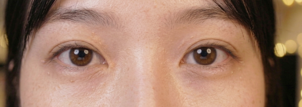
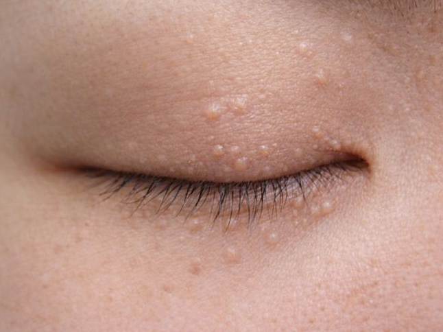
Many people come in saying, “I have these bumpy spots around my eyes, and I don’t know what they are.” If you look closely, they may appear as small white granules on the skin surface or as flesh-colored, raised papules.
Although these two conditions may look similar, their causes and locations of origin are completely different skin conditions. Today, we’ll take a close look at the difference between syringoma and milium, and review the proper way to respond.
Skin surface vs. dermis,
the location of onset is different from the start.
Milium occurs just below the epidermis,
that is, in the shallowest layer of the skin,
as a small cyst.
It usually appears as a white granule-like
hard mass when keratin gets trapped
inside the skin.
By contrast, syringoma is a benign tumor
that develops when the duct cells of
eccrine sweat glands in the dermis
proliferate excessively.
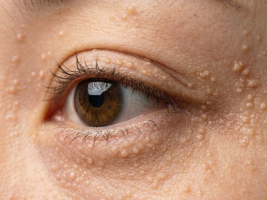
It is characterized by multiple 1–3 mm papules
that gather beneath the eyes, especially
around the orbit, and appear flesh-colored
or slightly yellowish.
The key to the difference between syringoma and milium
is this “depth.”
Because milium starts in the epidermis, while syringoma
starts in the dermis, even if both cause bumpy
symptoms in the eye area, the approach
may need to be different.

Why doesn’t dermatology recommend self-extraction?
When something small appears on the skin,
it is understandable to want to squeeze it
with your fingers or try to pop it with a pin.
However, syringoma in particular is located deep
within the dermis, so if you try to extract it
yourself, the lesion may not be removed well,
and the risk of wounds and inflammation on the
skin surface is high.
Even for milium, although it is in the epidermis,
unsanitary tools and excessive force can lead
to hyperpigmentation or scarring.
The skin around the eyes is very thin and sensitive,
so even minor irritation can take a long time
to heal.
Attempting self-extraction without accurately
understanding the difference between syringoma and milium
often ends up making the skin condition worse.
How does a CO2 laser work?
At Ijiham Dermatology,
CO2 lasers are used to manage
syringoma and milium.
A CO2 laser uses infrared light with a wavelength
of 10,600 nm, which is absorbed by water in the
skin tissue and works by precisely vaporizing
the lesion.
Because the lesion tissue can be treated
layer by layer with precision,
it is useful for reducing damage to the surrounding
normal tissue.
For milium, the keratin content and cyst wall of the epidermal cyst
are treated, and for syringoma, the approach is gradual
so that the energy can reach the tumor tissue
present in the dermis.
In particular, because syringoma is deeper,
rather than trying to remove it all at once,
it is important for scar prevention to divide the treatment
into stages while considering the skin’s recovery cycle.
Trying to remove everything at once can instead cause scars
There are cases we often see in the clinic.
Some patients want to get rid of eye-area syringoma
in a short period of time and ask for all lesions
to be treated in a single session.
However, the dermis around the eyes is very thin
and has abundant blood flow.
If excessive laser energy is concentrated over a short
period of time, the normal dermal tissue can be damaged,
and scars or pigment changes may remain.
Because the depth and characteristics of each lesion differ
depending on the difference between syringoma and milium,
a step-by-step approach becomes an important principle
for protecting the skin condition.
"The skin needs time to heal on its own. Even if it is a little slower,
respecting the skin’s recovery process can ultimately lead to
better changes."
This is the direction of care that Ijiham pursues.

How to care for the eye area after CO2 laser treatment
After CO2 laser treatment, a scab (crust) forms
on the treated area.
This scab is a natural part of the skin recovery process,
so it is important not to forcibly remove it.
For 1–2 weeks after treatment, avoid direct friction
on the area when washing your face, and be sure to
use sunscreen.
The area around the eyes is prone to pigmentation
caused by ultraviolet exposure, so we recommend
making SPF 50 or higher part of your daily routine.
Moisturizing is also important.
During the process of skin barrier recovery,
adequate hydration can help maintain the
rhythm of skin healing.
It is also best to avoid heat-stimulating environments
such as saunas or jjimjilbangs for one week
after treatment.

A clear diagnosis between syringoma and milium comes first
When bumpy spots appear around the eyes,
many people try to judge and handle them on
their own.
However, syringoma and milium are not easy to distinguish
with the naked eye, and it is not uncommon for both
to exist at the same time.
If you approach them in the same way without an accurate
diagnosis, treatment efficiency may be low or unnecessary
skin damage may occur.
Ijiham Dermatology practices throughout its care
the philosophy of “creating a comfortable face,
rather than excessive change.”
Rather than chasing noticeable change in a short time,
we value a process that follows the skin’s recovery rhythm
and moves forward step by step without scarring.
With honest care and appropriate methods,
we will work together to design a treatment plan that
fits the skin condition of each visitor.
We hope this article was helpful to those who were curious
about the difference between syringoma and milium.
Ijiham Dermatology will always provide honest care
from the same place.











 Previous image
Next image
Previous image
Next image









Previous image
Next image









Previous image
Next image