Hello. I’m Dr. Jo Hyun-woo of 입체성형외과.
When patients go to consultations for facial contouring surgery, they often get a CT scan.
Sometimes, while consulting, there are patients who can read the CT almost as well as a doctor.
So today, I’d like to organize and explain the things that are worth checking on a CT during a consultation.
The photo above is the 3D CT image you usually see first when going for a consultation.
What you should pay attention to in this image is how symmetrical your face is.
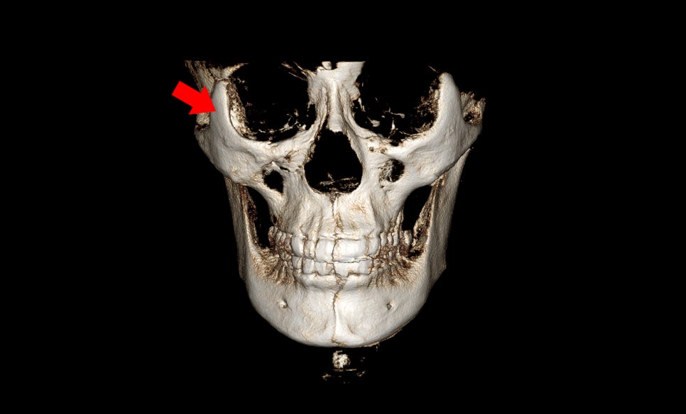
First, look at the position of the eye sockets.
If the left and right heights of the eye sockets are different, there will be a significant difference in the height of the cheekbones.
In such cases, correction of asymmetry may be necessary.
However, as I always say, because surgery to change the position of the eye sockets has limitations as a cosmetic procedure, complete correction of cheekbone asymmetry is not possible.
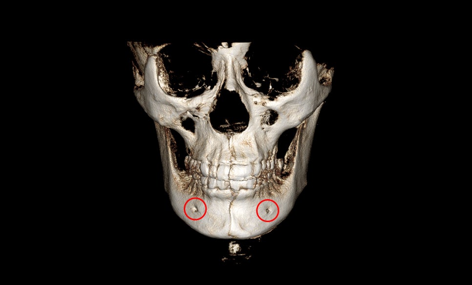
The second thing to look at is the location where the nerve of the lower jaw comes out.
In this patient, the position of the hole where the mandibular nerve line comes out is lower on the left side.
In this case, you can think of the left jawbone as protruding lower.
If you look at it this way, you can see that the left jawbone extends farther downward.
Therefore, you can identify asymmetry of the lower jawbone just by the location of the hole where the nerve line comes out.
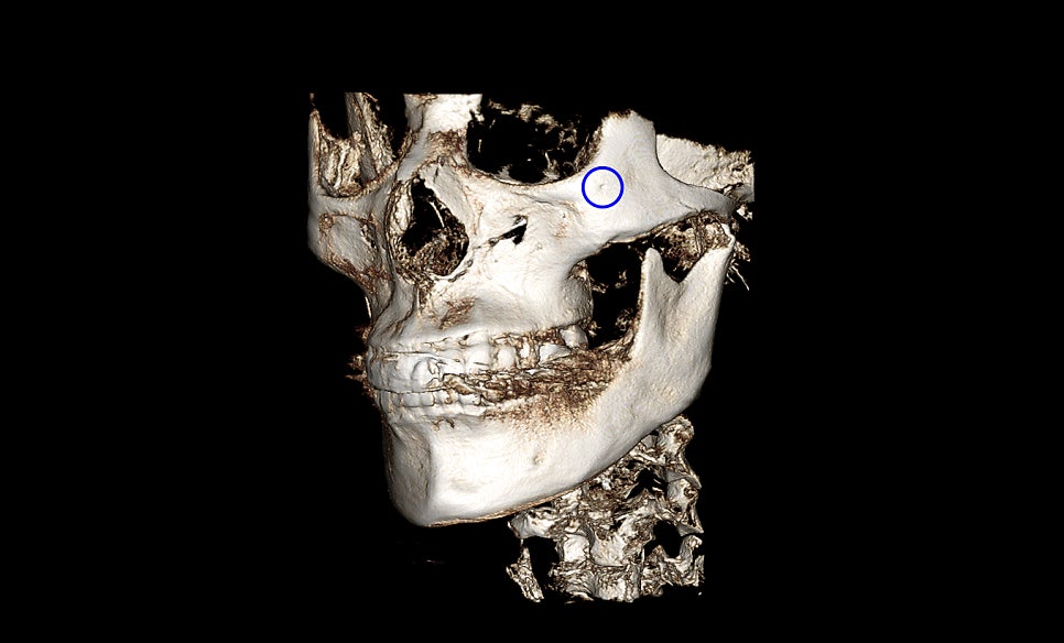
Next, you can look at the location of the “zygomaticotemporal nerve” on the outer side of the cheekbone.
The area marked with a blue circle in the illustration is the location where the nerve comes out.
If this location is close to the eye socket, more of the 45-degree cheekbone can be cut, and if it is located lower, it has to be cut more conservatively, so I think it is good to know this point as well.
Next, I will explain what can be seen on regular CT images rather than 3D CT.
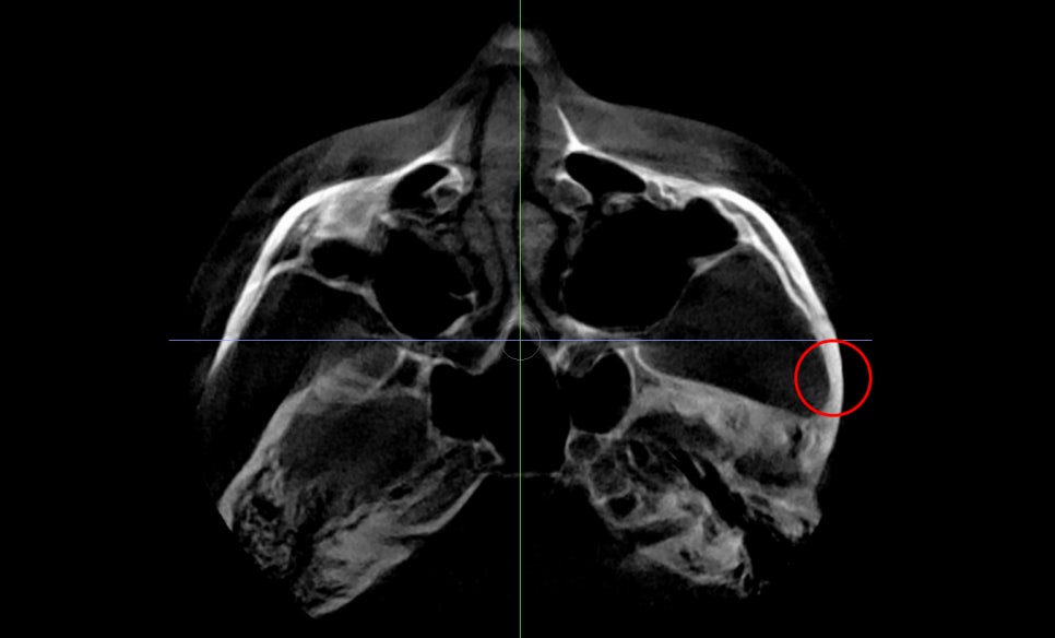
This is the image referred to as the axial view of the CT. In this image, you can see the thickness of the side cheekbone.
If the thickness of the marked bone is great, the side cheekbone can be reduced more. If the bone is thin, the amount that can be reduced is smaller, so it is also important to understand this thickness.
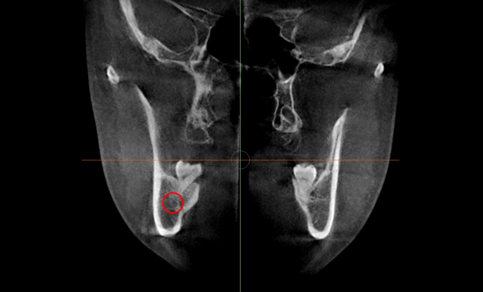
This is the coronal view of the CT. Here, when we perform lower jaw surgery, we can check how close the inferior alveolar nerve is to the cortex.
We can also check how much masseter muscle there is.
The structure visible on the outer side of the jawbone is the masseter muscle, and in order to improve the frontal effect, it is essential for this masseter muscle to become smaller.
Finally, I will show you the panoramic view.
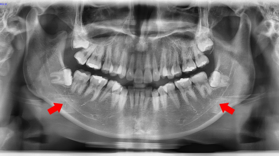
This is the image we often take at the dentist.
In this image, you can see the opening where the inferior alveolar nerve comes out of the lower jaw and its pathway.
The white line passing through like this is the path of the inferior alveolar nerve. The opening appears as a circle.
If the inferior alveolar nerve is high, more of the lower jaw can be cut, and if it is low, the amount that can be cut may be smaller, so knowing these points may be helpful when explaining the surgery.
So far, I have explained how to read CT images that may be helpful during a facial contouring surgery consultation.
Of course, you may not be able to read them as accurately as an expert, but if you understand these basic points, I think you will be able to understand and judge things more accurately.
Thank you.