Hello, this is Director Jo Hyun-woo of 입체성형외과.
These days, many patients come in for consultations and then take copies of their own CT images with them.
CT and panoramic images can be viewed accurately by experts, but today I’d like to explain a way for patients to understand CT images more easily.
In general, CT images are composed of files called Dicom.
If you put them on a USB drive, you can run them with Autorun and view them.
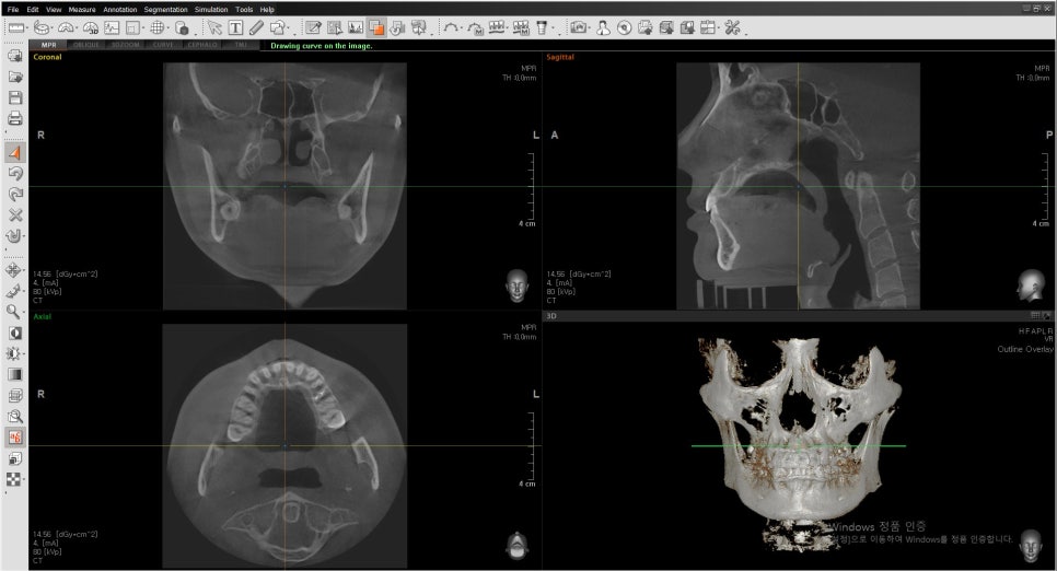
This screen is the first screen you can see when viewing a CT.
Then let’s look at what these four screens mean.
The easiest one to understand is the image at the bottom right.
This is a 3D CT image of your facial bones.
This 3D screen is not an actual 3D scan of the patient’s face; it is a screen created by computer software in three dimensions based on the image at the bottom left.
CT is a tomographic scan, and you can think of it as creating an image by overlapping multiple X-ray images.
Using X-rays, cross-sections of the body are captured.
This is called the axial view in CT. To put it simply, if you look at the diagram:
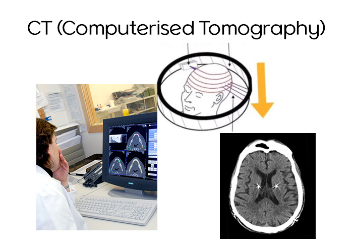
It means taking multiple sectional images like this.
When CT is first taken or set up, the spacing for these axial views is configured.
Of course, taking images at 1 mm intervals will provide a more accurate image, while taking them at 1 cm intervals can leave gaps in between.
You can think of those gaps as being smoothly reconstructed by computer software.
The better the CT, the narrower this interval is, so the image comes out more precisely.
In general, CT machines in plastic surgery clinics are dental CTs,
and those intervals are not that precise.
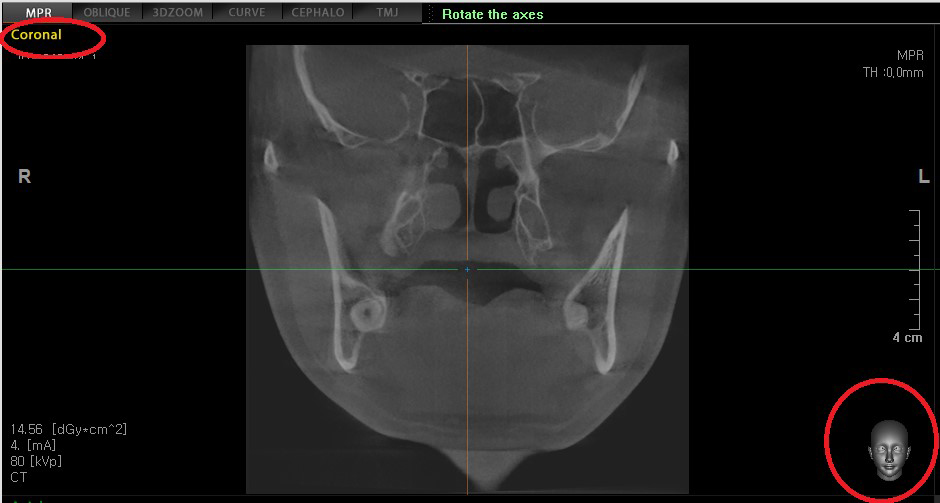
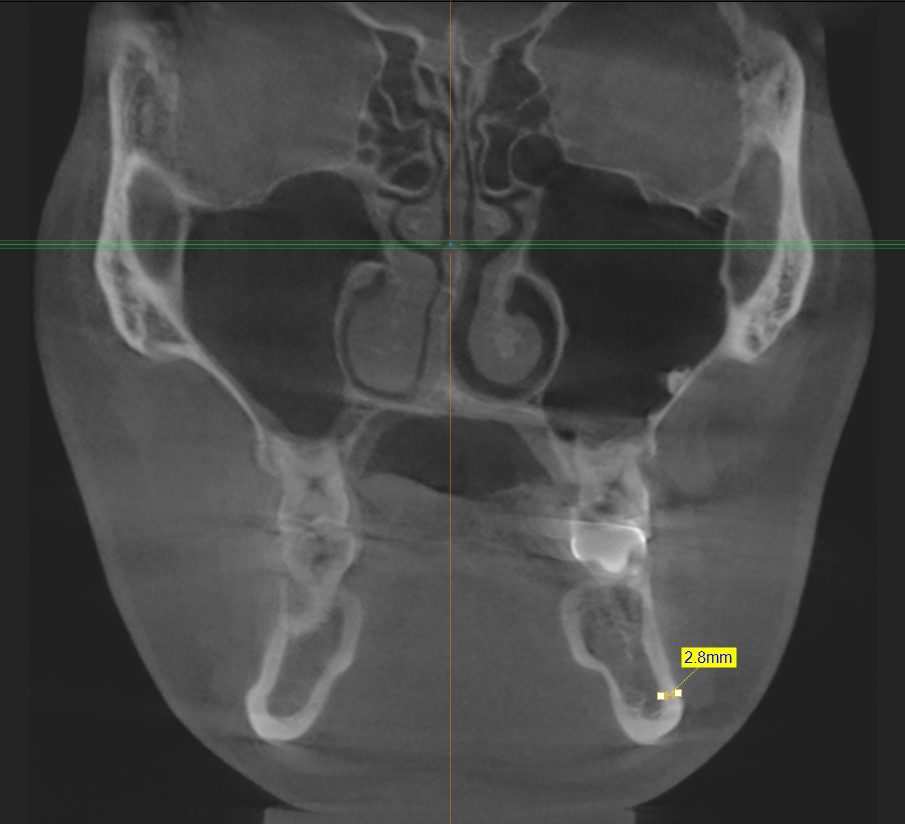
The image shown at the upper left is called the coronal view.
It is a view seen by slicing the image from the front, as shown in the lower right illustration.
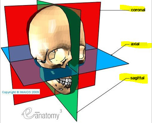
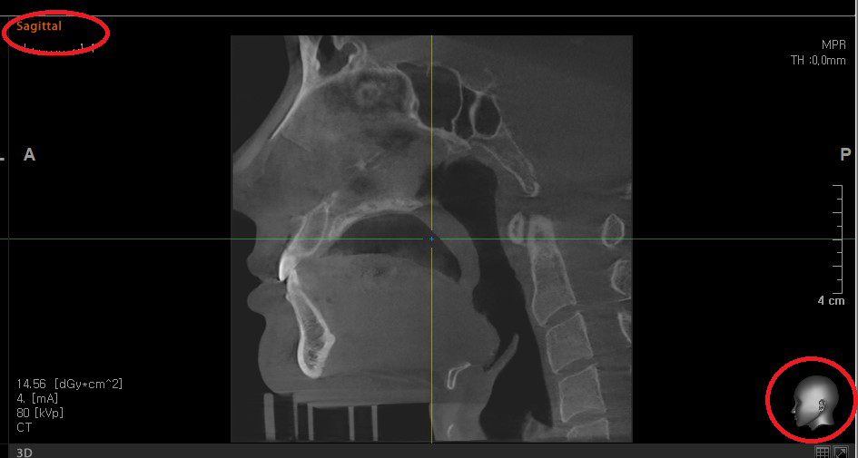
The image in the upper right is the sagittal view.
It may be easier to understand if you think of it as an image taken by cutting through the side profile.
Then what kinds of information can be easily obtained from a CT, which contains so much information?
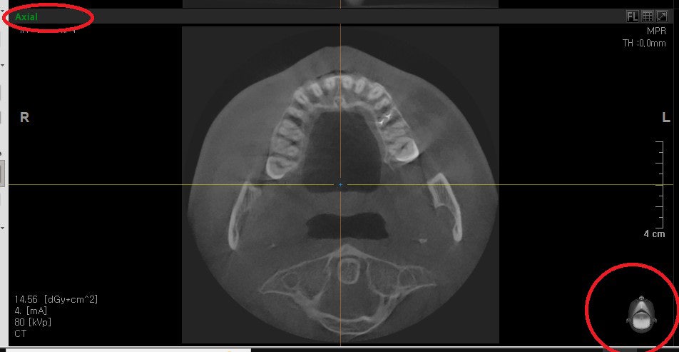
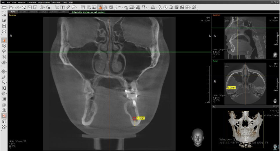
- First, how far can my cheekbones be moved inward?
This information can be checked in the axial view.
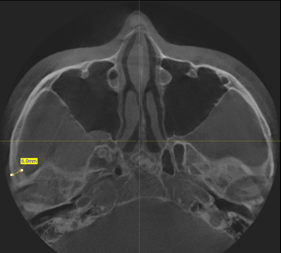
If you scroll up and down through the CT images and come across a view like this, stop and measure the thickness of the cheekbone.
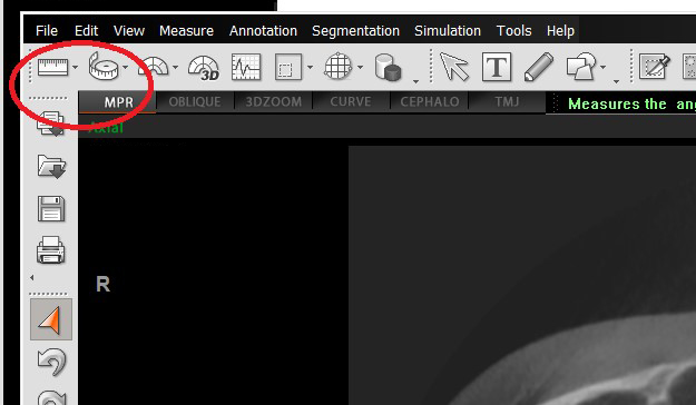
By clicking the ruler-like icon shown here, you can measure the thickness of your side cheekbone.
In this case, since the patient is a man, the thickness of the cheekbone on one side measures 6 mm.
Then you can infer that a total of 1.2 cm of bone can be moved inward on both sides.
- How much can I perform cortical osteotomy?
This information can be seen in the coronal view.
As you can see in the CT above, if you measure the thickness of the cortex, it is about 2.8 mm.
If this much cortical bone is removed, the face can appear noticeably slimmer from the front.
- How much of the square jaw bone can be removed?
This can also be seen in the coronal view.
If you look at the inside of the jawbone in the CT, you can see a round hole like this.
This hole is the passage through which the nerve runs.
If you actually measure the height from the very bottom of the bone, it comes out to 5.9 mm.
This image is taken when the nerve is passing through the lowest point.
If it is cut to the maximum, it can be cut by about 5.9 mm.
However, as I always say, if you cut too close, the nerve canal can become exposed, and even if the nerve itself is not damaged, touching that area after surgery may cause pain or persistent discomfort.
It is better to leave a margin of about 2–3 mm.
- Do I have more fat or more muscle?
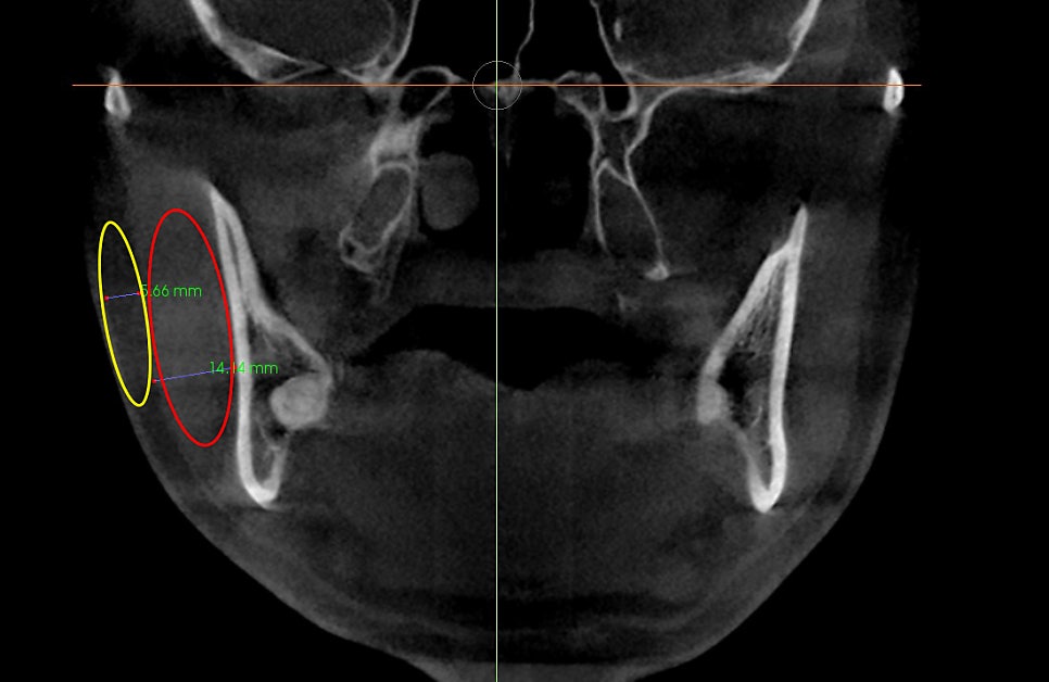
A lot of information can be obtained from the coronal view.
The light gray area outside the bone is muscle. The muscle thickness is listed as 14.1 mm.
The darker gray area outside that is the fat layer. It is 5.66 mm.
Of course, from these absolute values alone, we cannot say whether they are thick or thin, but we can still get a sense of the thickness to some extent.
Compared with the fat of average others, it is thicker, and the muscle is on the thinner side.
You can hear a more detailed comparison through a consultation.
- How much can my chin bone move forward or backward?
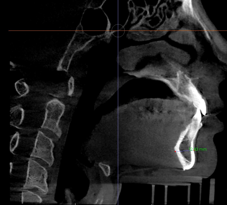
This information can be seen in the sagittal view.
If you stop at the area where the chin bone is visible and measure its front-to-back thickness, it comes out to about 12.4 mm.
In my case, when moving bone forward or backward, I perform surgery with the idea that the contact area of the bone should be at least about half so that the bone can heal well.
Therefore, the conclusion is that the bone can be moved forward or backward by about 6–7 mm.
For some people it is around 10 mm, and for others it can be up to 16 mm, so it would be good to make a judgment based on that.
So far, I’ve explained how to find five kinds of information.
In fact, there is more information in CT scans, but those are things that patients may find difficult to understand.
Just knowing these five points should be enough for you to understand your own bones well, and you may be able to understand them accurately without having to go back to the hospital for another explanation.
CT can have errors depending on its precision, and 3D images can also be inaccurate.
Even if something on a 3D CT looks like a nonunion, the bones may actually be fully joined, so you do not need to judge everything based only on the CT or worry too much.
Thank you.