Hello, I’m Dr. Jo Hyun-woo of Statura Plastic Surgery.
When patients go to facial contouring consultations or come in for follow-up visits, they often get CT scans. Sometimes, there are even patients who can read CT scans almost as well as doctors.
Today, I’d like to organize and explain the things you should definitely look at on a CT scan.
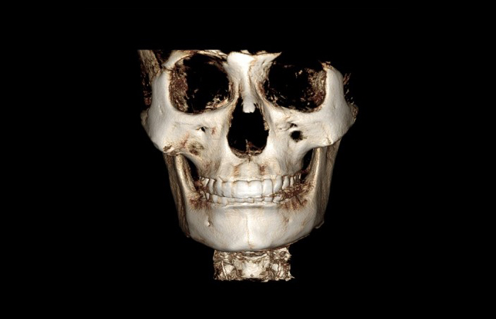
This is a CT image that is commonly seen for the first time during a consultation.
What you should pay attention to in this image is how symmetrical your face is.
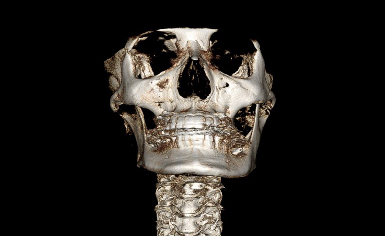
First, look at the position of the eye bones.
If the height of the eye bones differs, the height of the cheekbones will also differ significantly.
In such cases, there may be situations where asymmetry needs to be corrected.
However, as I always say, surgery to change the position of the eye bones has limitations as a cosmetic procedure, so complete correction of cheekbone asymmetry is not possible.
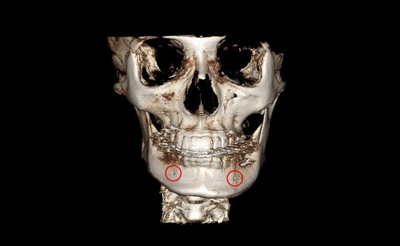
The second thing to look at is the location where the nerve of the lower jaw comes out.
In this patient’s image, the opening where the lower jaw nerve line emerges is lower on the left side.
In this case, you can think of the left jawbone as protruding further downward.
As you can see here, the left jawbone is positioned lower.
Therefore, just by looking at the position of the opening where the nerve exits, you can check asymmetry in the lower jawbone.
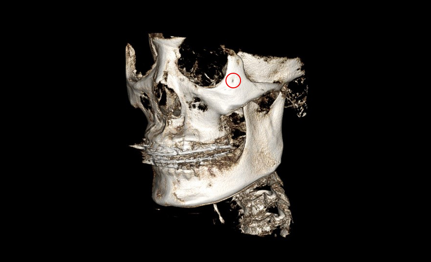
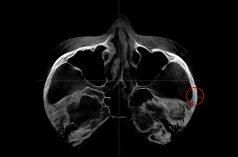
Next, you can look at the location of the zygomaticotemporal nerve in the cheekbone.
The part marked with a red circle in the illustration is where the nerve exits.
If this location is close to the eye bone, more of the 45-degree cheekbone can be removed. If it is located farther outward, it can only be removed more conservatively, so it may be helpful to know this point.
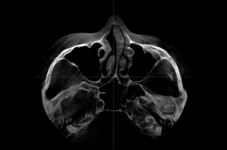
Next is the image referred to as the axial view on CT.
In this image, you can determine the thickness of the side cheekbone.
If the bone is thick, more of the lateral cheekbone can be reduced, and if the bone is thin, the amount that can be reduced is smaller, so it is important to understand this thickness.
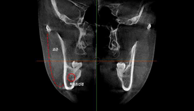
This image is the coronal view on CT.
Here, when we perform jaw surgery, we can check how close the inferior alveolar nerve is to the cortex.
We can also 확인 how much masseter muscle there is.
What appears on the outer side of the jawbone is the masseter muscle.
To improve the frontal effect, reducing this masseter muscle can be considered essential.
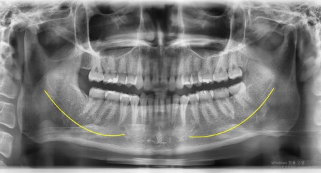
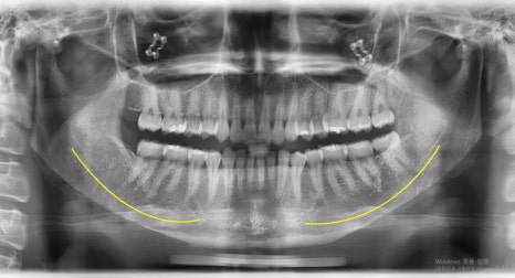
Lastly, let me show you the panoramic view. This is the type of image that is often taken at the dentist.
In this image, you can see the course of the inferior alveolar nerve in the lower jaw and the opening where it exits.
The yellow line shown here is the path along which the inferior alveolar nerve runs.
If the inferior alveolar nerve is high, more of the lower jaw can be trimmed, and if it is low, the amount removed may be smaller, so knowing these points may help you understand the surgical explanation.
So far, I’ve explained how to read CT images in ways that may be helpful during facial contouring consultations.
Of course, you may not be able to read them as accurately as a specialist, but if you understand these basic points, I think you will be able to understand and judge things more accurately.
Thank you.