Hello.
This is E-pyeonhansesang Dental Clinic.
Today, we will look at the importance of diagnosis before wisdom tooth extraction
and the inferior alveolar nerve located in the lower jaw.
Many patients say this:
"Do I really need an examination before extraction?"
"I already had an X-ray, so why do I need a CT scan?"
If you have had these questions,
please stay with us until the end of this post.
chapter. 1
Why is diagnosis needed before wisdom tooth extraction?
Wisdom teeth are called third molars,
and they are located at the very back of the mouth.
They are teeth with very complex relationships
with the jawbone, nerves, blood vessels, and adjacent teeth.
If you approach it simply as just pulling a tooth,
it may seem straightforward, but in fact there are countless
variables involved.
Therefore, if extraction begins without a diagnostic process,
unintended complications may occur.
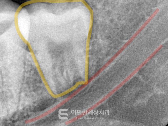
*Image for illustration purposes (inferior alveolar nerve: red solid line)
In particular, for lower wisdom teeth, a deep understanding
of the anatomical structure called the inferior alveolar nerve is needed.
In the example image, you can see a nerve strand
running long near the root.
This is the part called the inferior alveolar nerve.
If the root tip invades the nerve canal
or comes close to it, damage may occur during extraction,
leading to temporary or permanent nerve palsy symptoms.
Therefore, it is important to check this carefully
and make an extraction plan.
Also, the difficulty of wisdom tooth extraction
varies greatly from person to person.
Whether it is a simple extraction,
or a surgical procedure that requires an incision in the gum,
or whether part of the bone must be removed,
precise examination is needed in advance
to determine the level of difficulty.
By identifying these factors beforehand,
the extraction time can be reduced and the patient's burden
(bleeding, pain, swelling, etc.) can be minimized.
Lastly, pre-extraction examination is needed
to detect diseases or lesions caused by the wisdom tooth.
To check whether the wisdom tooth is pressing on the surrounding teeth,
whether root resorption has occurred,
whether there is a cyst at the root,
or the condition of the gum bone,
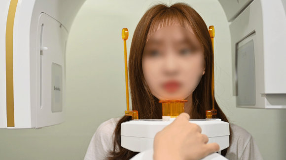
a pre-extraction X-ray and CT scan are performed.
| 🦷 Wisdom Tooth Encyclopedia Summary Why is an examination needed before wisdom tooth extraction? |
|---|
| ☑️ To reduce the risk of nerve (inferior alveolar nerve) damage |
| ☑️ To predict the difficulty of extraction |
| ☑️ To check for accompanying diseases and the condition of surrounding structures |
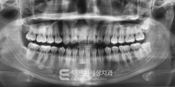
*Image for illustration purposes. (Panoramic image)
chapter. 2
Why is CT additionally taken?
When extracting a wisdom tooth, the most basic
imaging used for diagnosis is an X-RAY.
This is called a panoramic radiograph,
and it is a very useful initial diagnostic method.
It is an important diagnostic method that allows you to see
at a glance the overall structure of the dentition, bone structure,
relationship with adjacent teeth, and the approximate root shape.
It also has the advantages of relatively low cost,
low radiation exposure, and simple imaging.
However, there are parts that are difficult to confirm
with a panoramic image alone.
Because panoramic imaging is a two-dimensional, partial diagnostic method,
there is some lack of depth, three-dimensionality,
and accurate distance information.
For example, if the root and nerve canal overlap
in a panoramic image,
it is difficult to determine the actual distance.
So, depending on the situation, a CT scan
is additionally performed.
CT allows us to check three-dimensional space.
Therefore, if the root crosses the nerve canal
or is adjacent to it on the panoramic image,
a CT scan is used to examine this area
in more detail.
A CT scan does not only allow us to check
the distance to the nerve canal.
It also allows detailed observation of the wisdom tooth root shape.
If the root is severely curved, thin,
or deeply embedded within the bone,
CT can also help determine the overall structure.
| 🦷 Wisdom Tooth Encyclopedia Summary Why is CT additionally taken? |
|---|
| ☑️ To check the close distance between the root and the inferior alveolar nerve |
| ☑️ To examine the root and shape of the wisdom tooth in detail |
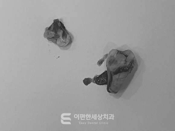
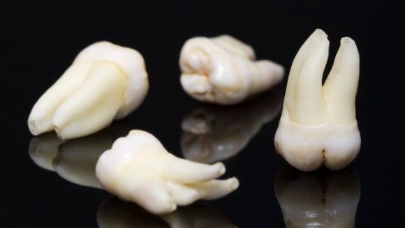
*Image for illustration purposes. (Extracted wisdom tooth)
chapter. 3
What is the wisdom tooth extraction process after diagnosis?
Then, once the pre-extraction diagnosis is complete,
what process is followed to finish the extraction?
If the patient's oral condition has been checked
through accurate diagnosis and clinical examination,
the actual extraction will begin.
(*Clinical examination refers to visual inspection and palpation of the oral cavity.)
After preparing the necessary tools for extraction,
the extraction method and access approach are planned.
Then, after anesthesia reduces sensation,
if necessary, the gum is incised and part of the bone is removed.
After that, when the wisdom tooth is visible,
it is removed from the gum and extracted,
and the gum is sutured to complete the procedure.
| 🦷 Wisdom Tooth Encyclopedia Summary What is the wisdom tooth extraction process? |
|---|
| 1️⃣ Diagnosis |
| 2️⃣ Extraction plan and preparation |
| 3️⃣ Anesthesia |
| 4️⃣ Extraction |
| 5️⃣ Suturing and completion |
Pre-extraction diagnosis for wisdom teeth
is not an option, but a necessity.
Through diagnosis, unexpected complications such as nerve damage can be prevented,
and a plan suitable for the level of extraction difficulty can be made.
Thank you.
[E-pyeonhansesang Dental Clinic Encyclopedia]
About precise diagnosis before wisdom tooth extraction