Hello,
I am Seo Ho-yeon, the chief director of Yonsei Dagam Dental Clinic.
I have been keeping a journal for 16 years now,
but this is my first time writing a blog, so I would appreciate it if readers would look kindly on it :)
Going forward, through this blog,
I will do my best to provide many people with
objective medical information.
The topic I have chosen for today’s first post after much thought is
“impacted supernumerary teeth.”
First, the term itself may be unfamiliar,
so I will explain the definition step by step.
In academic terminology,
it is called a “supernumerary tooth.”
It refers to teeth that have erupted in addition to the normal number.
Source: Google
Since it has formed beyond the normal number,
there is often not enough space,
so it is common for it to remain
unable to erupt inside the jawbone.
This is called an “impacted supernumerary tooth.”
If its position is unfavorable and it disrupts the dental arch,
or if it causes resorption of the roots of nearby adjacent teeth,
it usually needs to be removed.
Example case
From here on, I will use a case from
Yonsei Dagam Dental Clinic to help with understanding.
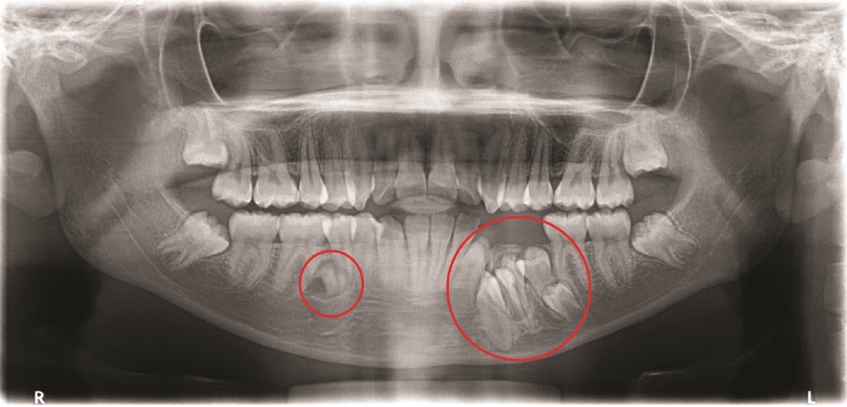
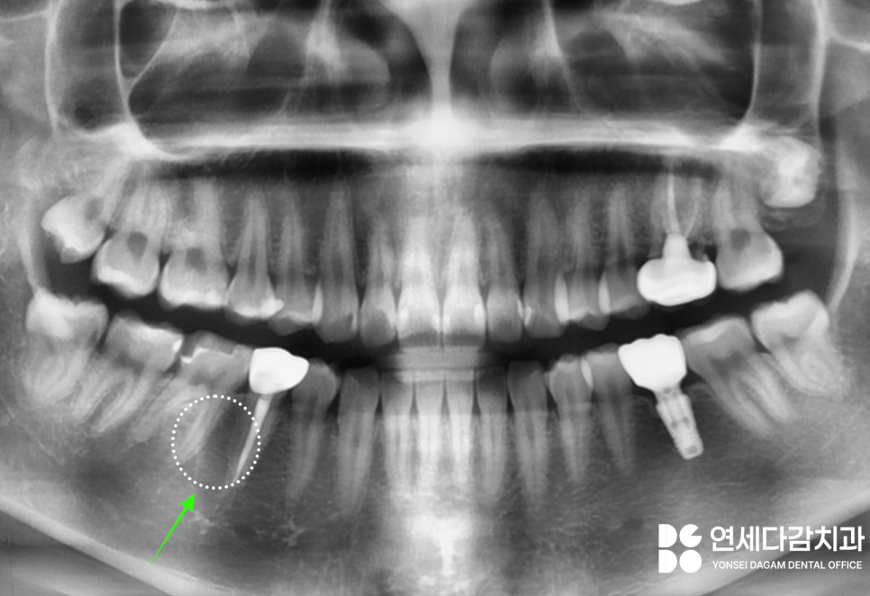
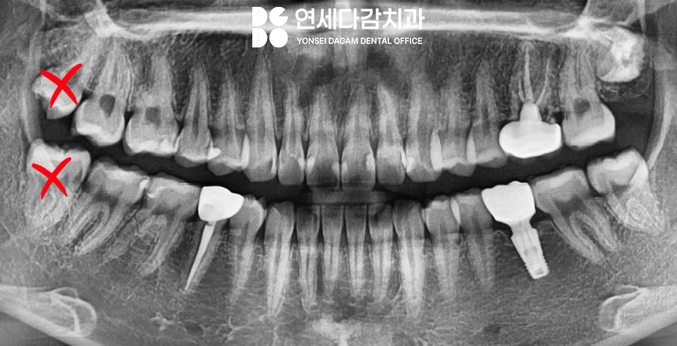
Initial X-ray (2023.12.18)
An impacted supernumerary tooth is present
in the lower right premolar area.
(It is usually first found on a panoramic X-ray.)
Even if there are no special symptoms,
considering various risk factors such as future cyst formation and root resorption of adjacent teeth,
removal is generally recommended.
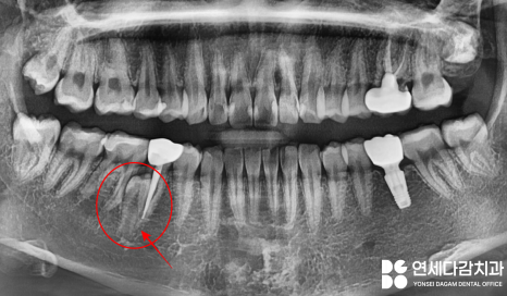
It had remained buried on the lingual side,
but the issue here was that it was located fairly close to the lingual nerve,
which is responsible for sensation in the tongue.
※ When extracting wisdom teeth, too,
we usually preserve the bone on the tongue side as much as possible,
because the lingual nerve is present along the bone wall on that side.
2023.12.18
2023.12.18
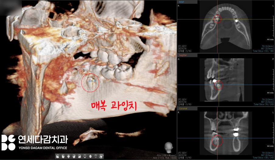
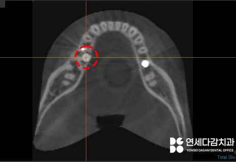
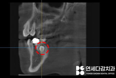
This is the CT scan taken before surgery at Yonsei Dagam Dental Clinic.
In addition to the 2D panoramic image,
we accurately identify the degree of impaction and its position through 3D CT,
and then carefully remove it so that surrounding structures are not damaged.
After identifying the exact location of the supernumerary tooth,
we incised the gingiva on the lingual side and completed the extraction on the same day.
In this case, fortunately,
there was a certain distance from the lingual nerve,
but
when removing an impacted supernumerary tooth on the lingual side,
it is necessary to be mindful of the lingual nerve
while performing mucosal retraction and extraction.
The next day, disinfection was performed.
2023.12.19
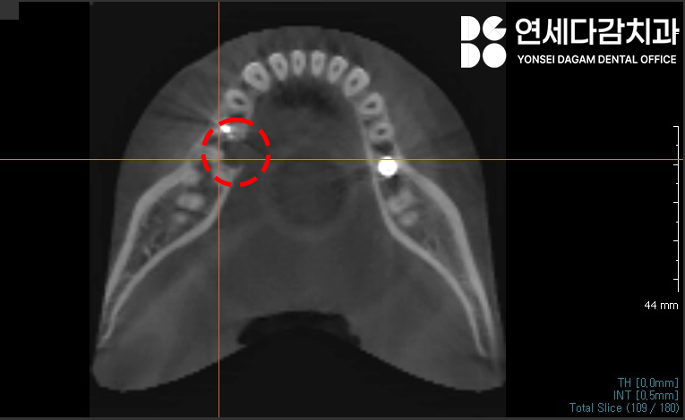
This is an X-ray and CT scan taken once more to check the surgical site along with disinfection.
You can see that it is healing well
without any particular abnormalities.
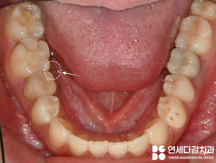
The photo below is an oral photograph from Yonsei Dagam Dental Clinic
taken 8 months after the procedure,
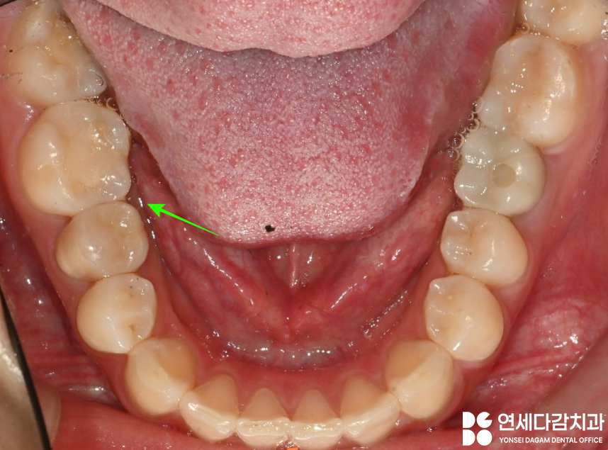
2024.08.19
and once again, you can confirm that the gums have healed well.
2024.08.19
*The right upper and lower wisdom teeth (#18, 48),
which were also difficult to manage later because of their position,
were removed together.
▶Treatment period: 2023.12.18~2023.12.28 (same day / disinfection the next day / suture removal 10 days later)
As in the extraction case from
Yonsei Dagam Dental Clinic that I showed today,
there are several structures that must be approached with caution
when extracting lower teeth.
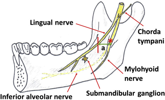
Source: https://www.sciencedirect.com/
(e.g., lingual nerve, inferior alveolar nerve canal, etc.)
Therefore,
if the position is judged to be unfavorable on a panoramic X-ray,
it is important to establish a treatment plan
along with a CT scan.
The more precise the diagnosis,
the more helpful it is in advance for understanding the extraction method,
time required, and anticipated complications.
That concludes the information prepared today by
Yonsei Dagam Dental Clinic.
Thank you ^^