Hello.
This is Yonsei Dagam Dental Clinic near Police Hospital Station.
Did you know that if a tooth is missing,
the bone can gradually be lost?
The alveolar bone is the bone that supports the teeth,
and it receives force during chewing
and plays an important role in
transmitting that force to the bone tissue.
Therefore, teeth and the jawbone
are closely connected to each other,
and if a tooth is missing, the
mechanical stimulation that maintains the bone disappears,
causing bone loss to begin.
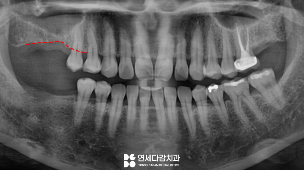
This is a case in which the missing teeth
in the upper and lower molar areas on the right side
were left untreated and then implants were performed.
Because it had been left for a long time,
the gum bone that had not received stimulation was resorbed,
and you can see a sunken appearance.
For an accurate diagnosis, a CT scan
was taken and reviewed,
and pneumatization of the maxillary sinus
was found.
What is pneumatization?
At the Police Hospital Station Dental Clinic,
we will explain it in detail.
First, the maxillary sinus is one of the sinuses,
located below the eyes near the cheekbone and close to
the root area of the upper molars,
and it is an air-filled cavity
covered with mucosa.
Pneumatization means that the base of the maxillary sinus
expands downward and invades the surrounding structures,
increasing the air-filled space.
This can gradually expand
with aging,
and if chronic inflammation persists, it can expand abnormally beyond that.
When that happens,
it resorbs the alveolar bone
and reduces the remaining bone height.
Therefore, the vertical bone height
is currently quite insufficient.
Placing implants in thin and lowered jawbone
comes with risk factors.
If surgery is performed in an environment
without enough bone volume,
the mucosa may be damaged by the instrument
and perforated.
It is best to avoid situations like this if possible.
So how was it solved at the Police Hospital Station Dental Clinic?
The method used is to lift the maxillary sinus,
expand the space, and then reinforce that space with bone graft material,
so that the fixture can be
securely fixed in place.
There are two approaches,
depending on the height of the bone.
Crestal Sinus Lift
This is called the crestal approach,
and it is a method of approaching from the site where the implant will be placed
and lifting the floor of the maxillary sinus
from below.
Since there is still some remaining bone,
it may be performed together with sinus lifting
and fixture placement.
If the bone is severely insufficient,
we proceed with the following method explained by the Police Hospital Station Dental Clinic.
Lateral Approach
This is a lateral approach used when the bone height is significantly lacking.
The gum is incised and the side of the bone is approached,
lifting the membrane while viewing it directly.
In the space created that way,
guided bone regeneration is performed.
After waiting until the reconstructed bone graft material stabilizes,
the implant is placed separately.
This method has the advantage of allowing the membrane to be directly viewed and manipulated,
but the recovery time may be somewhat longer.
To summarize,
if primary stability can be maintained,
it can be performed at the same time as placement,
but in cases where the vertical bone is insufficient, it is better to secure
primary stability,
so it is safer to perform the bone graft first,
allow healing, and then place the implant.
The diagnosis at the Police Hospital Station Dental Clinic
showed that the crestal approach would be possible.
Unless the vertical bone volume is severely insufficient,
using the crestal approach can shorten the healing process,
which may be less difficult for the patient.
This method is divided into two types.
✔ Hydraulic sinus lift method.
✔ A method of pushing the bone up using a special surgical kit.
Among the two methods above,
the hydraulic sinus lift method was used.
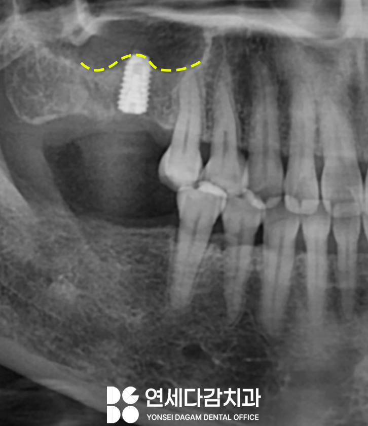
It was confirmed through X-ray that the existing vertical bone,
which was less than 3 mm,
increased to about three times more after lifting.
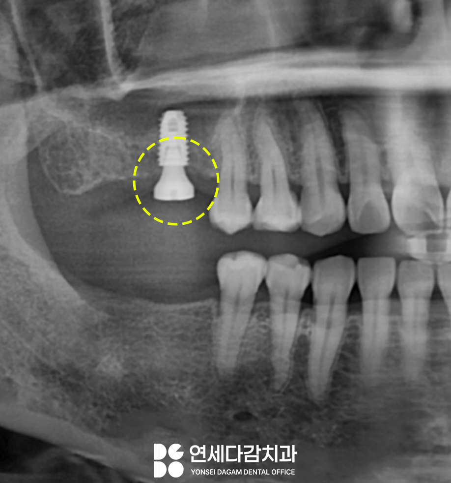
After about 3 months of osseointegration,
a healing abutment was connected.
This plays a role in creating the appropriate gum shape
to allow the prosthesis to be stably mounted.
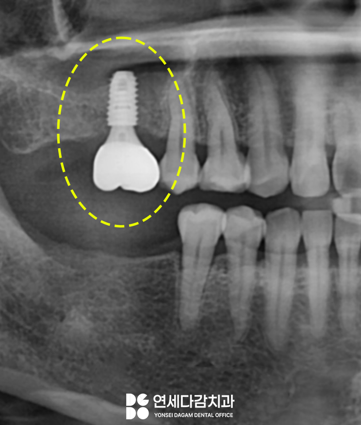
After about 2 weeks of soft tissue healing,
the final prosthesis was fabricated
and placement was completed.
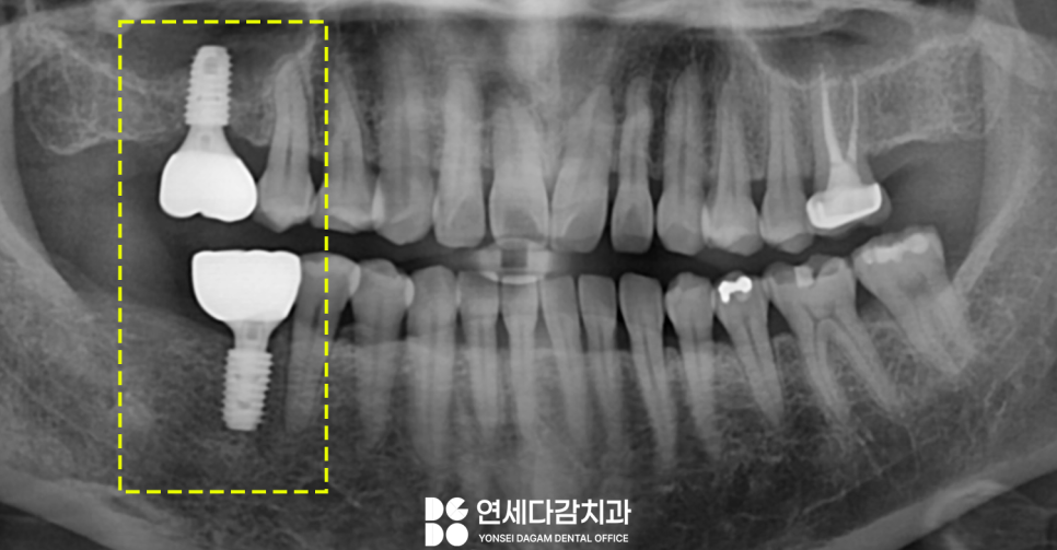
The missing space on the opposite side
was treated with simple implant placement,
and treatment was completed in both the upper and lower molar areas.
As explained today by the Police Hospital Station Dental Clinic,
if a tooth is extracted and left alone,
the alveolar bone gradually resorbs
and can cause various problems.
This is not simply due to aging,
but is a natural process that occurs for physiological reasons such as
✔ loss of mechanical stimulation
✔ loss of the periodontal ligament
✔ activation of osteoclasts
Therefore, if you are in a situation where a tooth must be extracted,
it is a good idea to prepare a plan for the recovery method afterward as well.
Through an accurate examination,
please check your individual oral condition
and consider the appropriate treatment method.
This was Yonsei Dagam Dental Clinic near Police Hospital Station.
Thank you!
Treatment period 2022.11.2-2023.5.10